Sodium »
PDB 3fak-3fvi »
3fbx »
Sodium in PDB 3fbx: Crystal Structure of the Lysosomal 66.3 kDa Protein From Mouse Solved By S-Sad
Protein crystallography data
The structure of Crystal Structure of the Lysosomal 66.3 kDa Protein From Mouse Solved By S-Sad, PDB code: 3fbx
was solved by
K.Lakomek,
A.Dickmanns,
U.Mueller,
R.Ficner,
with X-Ray Crystallography technique. A brief refinement statistics is given in the table below:
| Resolution Low / High (Å) | 32.11 / 2.40 |
| Space group | C 1 2 1 |
| Cell size a, b, c (Å), α, β, γ (°) | 148.804, 89.672, 64.954, 90.00, 98.67, 90.00 |
| R / Rfree (%) | 15.6 / 19.8 |
Other elements in 3fbx:
The structure of Crystal Structure of the Lysosomal 66.3 kDa Protein From Mouse Solved By S-Sad also contains other interesting chemical elements:
| Xenon | (Xe) | 1 atom |
Sodium Binding Sites:
The binding sites of Sodium atom in the Crystal Structure of the Lysosomal 66.3 kDa Protein From Mouse Solved By S-Sad
(pdb code 3fbx). This binding sites where shown within
5.0 Angstroms radius around Sodium atom.
In total only one binding site of Sodium was determined in the Crystal Structure of the Lysosomal 66.3 kDa Protein From Mouse Solved By S-Sad, PDB code: 3fbx:
In total only one binding site of Sodium was determined in the Crystal Structure of the Lysosomal 66.3 kDa Protein From Mouse Solved By S-Sad, PDB code: 3fbx:
Sodium binding site 1 out of 1 in 3fbx
Go back to
Sodium binding site 1 out
of 1 in the Crystal Structure of the Lysosomal 66.3 kDa Protein From Mouse Solved By S-Sad
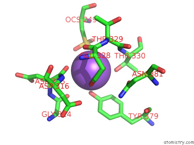
Mono view
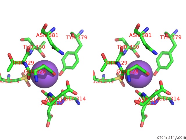
Stereo pair view
Mono view
Stereo pair view
A full contact list of Sodium with other atoms in the Na binding
site number 1 of Crystal Structure of the Lysosomal 66.3 kDa Protein From Mouse Solved By S-Sad within 5.0Å range:
|
Reference:
K.Lakomek,
A.Dickmanns,
U.Mueller,
K.Kollmann,
F.Deuschl,
A.Berndt,
T.Lubke,
R.Ficner.
De Novo Sulfur Sad Phasing of the Lysosomal 66.3 kDa Protein From Mouse Acta Crystallogr.,Sect.D V. 65 220 2009.
ISSN: ISSN 0907-4449
PubMed: 19237744
DOI: 10.1107/S0907444908041814
Page generated: Mon Oct 7 09:00:25 2024
ISSN: ISSN 0907-4449
PubMed: 19237744
DOI: 10.1107/S0907444908041814
Last articles
Cl in 5QF7Cl in 5QED
Cl in 5QFA
Cl in 5QEW
Cl in 5QEJ
Cl in 5QEV
Cl in 5QES
Cl in 5QEU
Cl in 5QDH
Cl in 5QEB