Sodium »
PDB 3epz-3f9l »
3f47 »
Sodium in PDB 3f47: The Crystal Structure of [Fe]-Hydrogenase (Hmd) Holoenzyme From Methanocaldococcus Jannaschii
Enzymatic activity of The Crystal Structure of [Fe]-Hydrogenase (Hmd) Holoenzyme From Methanocaldococcus Jannaschii
All present enzymatic activity of The Crystal Structure of [Fe]-Hydrogenase (Hmd) Holoenzyme From Methanocaldococcus Jannaschii:
1.12.98.2;
1.12.98.2;
Protein crystallography data
The structure of The Crystal Structure of [Fe]-Hydrogenase (Hmd) Holoenzyme From Methanocaldococcus Jannaschii, PDB code: 3f47
was solved by
T.Hiromoto,
O.Pilak,
E.Warkentin,
R.K.Thauer,
S.Shima,
U.Ermler,
with X-Ray Crystallography technique. A brief refinement statistics is given in the table below:
| Resolution Low / High (Å) | 25.00 / 1.75 |
| Space group | I 41 2 2 |
| Cell size a, b, c (Å), α, β, γ (°) | 95.930, 95.930, 165.810, 90.00, 90.00, 90.00 |
| R / Rfree (%) | 17.3 / 20.5 |
Other elements in 3f47:
The structure of The Crystal Structure of [Fe]-Hydrogenase (Hmd) Holoenzyme From Methanocaldococcus Jannaschii also contains other interesting chemical elements:
| Iron | (Fe) | 1 atom |
Sodium Binding Sites:
The binding sites of Sodium atom in the The Crystal Structure of [Fe]-Hydrogenase (Hmd) Holoenzyme From Methanocaldococcus Jannaschii
(pdb code 3f47). This binding sites where shown within
5.0 Angstroms radius around Sodium atom.
In total only one binding site of Sodium was determined in the The Crystal Structure of [Fe]-Hydrogenase (Hmd) Holoenzyme From Methanocaldococcus Jannaschii, PDB code: 3f47:
In total only one binding site of Sodium was determined in the The Crystal Structure of [Fe]-Hydrogenase (Hmd) Holoenzyme From Methanocaldococcus Jannaschii, PDB code: 3f47:
Sodium binding site 1 out of 1 in 3f47
Go back to
Sodium binding site 1 out
of 1 in the The Crystal Structure of [Fe]-Hydrogenase (Hmd) Holoenzyme From Methanocaldococcus Jannaschii
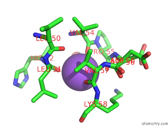
Mono view
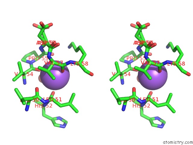
Stereo pair view
Mono view
Stereo pair view
A full contact list of Sodium with other atoms in the Na binding
site number 1 of The Crystal Structure of [Fe]-Hydrogenase (Hmd) Holoenzyme From Methanocaldococcus Jannaschii within 5.0Å range:
|
Reference:
T.Hiromoto,
K.Ataka,
O.Pilak,
S.Vogt,
M.S.Stagni,
W.Meyer-Klaucke,
E.Warkentin,
R.K.Thauer,
S.Shima,
U.Ermler.
The Crystal Structure of C176A Mutated [Fe]-Hydrogenase Suggests An Acyl-Iron Ligation in the Active Site Iron Complex. Febs Lett. V. 583 585 2009.
ISSN: ISSN 0014-5793
PubMed: 19162018
DOI: 10.1016/J.FEBSLET.2009.01.017
Page generated: Mon Oct 7 08:56:39 2024
ISSN: ISSN 0014-5793
PubMed: 19162018
DOI: 10.1016/J.FEBSLET.2009.01.017
Last articles
Cl in 5FQLCl in 5FRD
Cl in 5FRI
Cl in 5FP6
Cl in 5FOQ
Cl in 5FQ9
Cl in 5FOB
Cl in 5FOO
Cl in 5FLY
Cl in 5FOM