Sodium »
PDB 3epz-3f9l »
3f3e »
Sodium in PDB 3f3e: Crystal Structure of Leut Bound to L-Leucine (30 Mm) and Sodium
Protein crystallography data
The structure of Crystal Structure of Leut Bound to L-Leucine (30 Mm) and Sodium, PDB code: 3f3e
was solved by
S.K.Singh,
C.L.Piscitelli,
A.Yamashita,
E.Gouaux,
with X-Ray Crystallography technique. A brief refinement statistics is given in the table below:
| Resolution Low / High (Å) | 44.44 / 1.80 |
| Space group | C 1 2 1 |
| Cell size a, b, c (Å), α, β, γ (°) | 89.361, 86.664, 81.382, 90.00, 95.94, 90.00 |
| R / Rfree (%) | 18 / 20.5 |
Sodium Binding Sites:
The binding sites of Sodium atom in the Crystal Structure of Leut Bound to L-Leucine (30 Mm) and Sodium
(pdb code 3f3e). This binding sites where shown within
5.0 Angstroms radius around Sodium atom.
In total 2 binding sites of Sodium where determined in the Crystal Structure of Leut Bound to L-Leucine (30 Mm) and Sodium, PDB code: 3f3e:
Jump to Sodium binding site number: 1; 2;
In total 2 binding sites of Sodium where determined in the Crystal Structure of Leut Bound to L-Leucine (30 Mm) and Sodium, PDB code: 3f3e:
Jump to Sodium binding site number: 1; 2;
Sodium binding site 1 out of 2 in 3f3e
Go back to
Sodium binding site 1 out
of 2 in the Crystal Structure of Leut Bound to L-Leucine (30 Mm) and Sodium
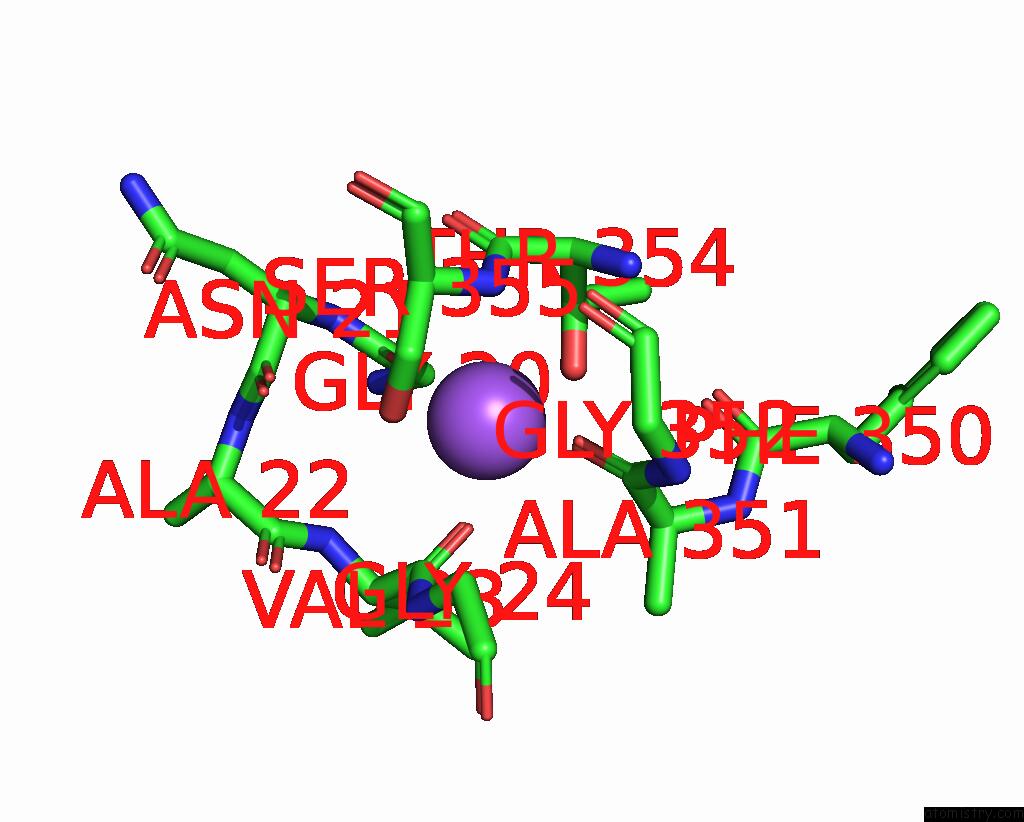
Mono view
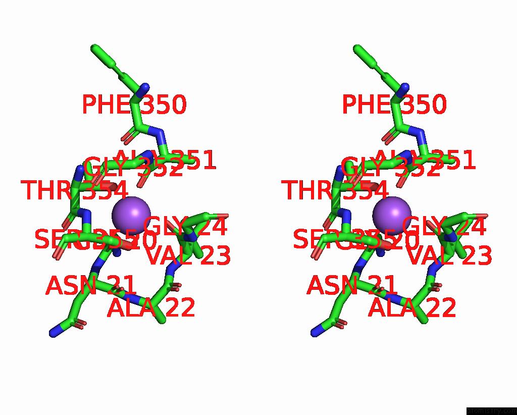
Stereo pair view
Mono view
Stereo pair view
A full contact list of Sodium with other atoms in the Na binding
site number 1 of Crystal Structure of Leut Bound to L-Leucine (30 Mm) and Sodium within 5.0Å range:
|
Sodium binding site 2 out of 2 in 3f3e
Go back to
Sodium binding site 2 out
of 2 in the Crystal Structure of Leut Bound to L-Leucine (30 Mm) and Sodium
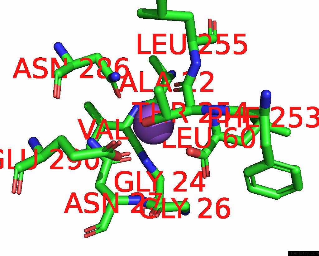
Mono view
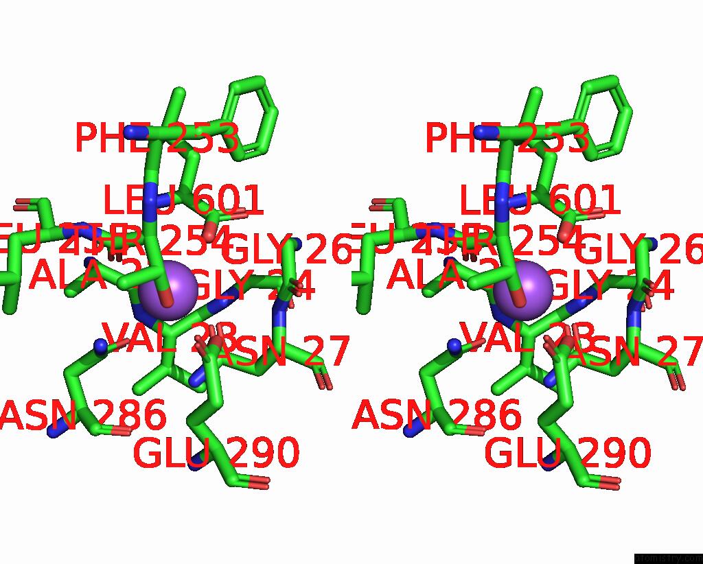
Stereo pair view
Mono view
Stereo pair view
A full contact list of Sodium with other atoms in the Na binding
site number 2 of Crystal Structure of Leut Bound to L-Leucine (30 Mm) and Sodium within 5.0Å range:
|
Reference:
S.K.Singh,
C.L.Piscitelli,
A.Yamashita,
E.Gouaux.
A Competitive Inhibitor Traps Leut in An Open-to-Out Conformation. Science V. 322 1655 2008.
ISSN: ISSN 0036-8075
PubMed: 19074341
DOI: 10.1126/SCIENCE.1166777
Page generated: Mon Oct 7 08:56:10 2024
ISSN: ISSN 0036-8075
PubMed: 19074341
DOI: 10.1126/SCIENCE.1166777
Last articles
Cl in 5FBWCl in 5FBV
Cl in 5FBU
Cl in 5FBN
Cl in 5FBT
Cl in 5FBR
Cl in 5FBQ
Cl in 5FAJ
Cl in 5FBL
Cl in 5FBH