Sodium »
PDB 3a07-3ahi »
3ahd »
Sodium in PDB 3ahd: Phosphoketolase From Bifidobacterium Breve Complexed with 2-Acetyl- Thiamine Diphosphate
Enzymatic activity of Phosphoketolase From Bifidobacterium Breve Complexed with 2-Acetyl- Thiamine Diphosphate
All present enzymatic activity of Phosphoketolase From Bifidobacterium Breve Complexed with 2-Acetyl- Thiamine Diphosphate:
4.1.2.22;
4.1.2.22;
Protein crystallography data
The structure of Phosphoketolase From Bifidobacterium Breve Complexed with 2-Acetyl- Thiamine Diphosphate, PDB code: 3ahd
was solved by
R.Suzuki,
T.Katayama,
B.-J.Kim,
T.Wakagi,
H.Shoun,
H.Ashida,
K.Yamamoto,
S.Fushinobu,
with X-Ray Crystallography technique. A brief refinement statistics is given in the table below:
| Resolution Low / High (Å) | 34.12 / 1.90 |
| Space group | I 4 2 2 |
| Cell size a, b, c (Å), α, β, γ (°) | 174.448, 174.448, 163.847, 90.00, 90.00, 90.00 |
| R / Rfree (%) | 16.2 / 19.7 |
Other elements in 3ahd:
The structure of Phosphoketolase From Bifidobacterium Breve Complexed with 2-Acetyl- Thiamine Diphosphate also contains other interesting chemical elements:
| Magnesium | (Mg) | 1 atom |
Sodium Binding Sites:
The binding sites of Sodium atom in the Phosphoketolase From Bifidobacterium Breve Complexed with 2-Acetyl- Thiamine Diphosphate
(pdb code 3ahd). This binding sites where shown within
5.0 Angstroms radius around Sodium atom.
In total only one binding site of Sodium was determined in the Phosphoketolase From Bifidobacterium Breve Complexed with 2-Acetyl- Thiamine Diphosphate, PDB code: 3ahd:
In total only one binding site of Sodium was determined in the Phosphoketolase From Bifidobacterium Breve Complexed with 2-Acetyl- Thiamine Diphosphate, PDB code: 3ahd:
Sodium binding site 1 out of 1 in 3ahd
Go back to
Sodium binding site 1 out
of 1 in the Phosphoketolase From Bifidobacterium Breve Complexed with 2-Acetyl- Thiamine Diphosphate
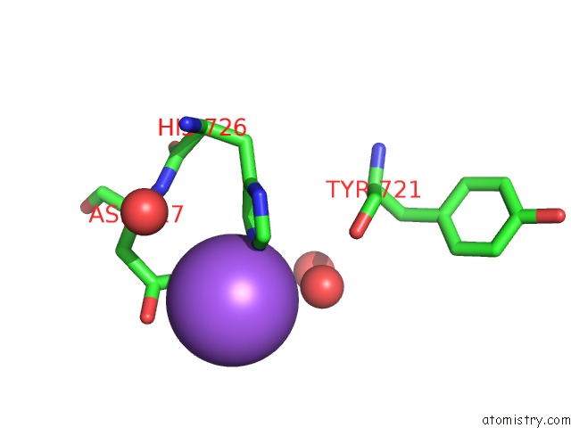
Mono view
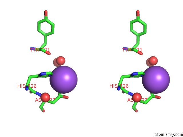
Stereo pair view
Mono view
Stereo pair view
A full contact list of Sodium with other atoms in the Na binding
site number 1 of Phosphoketolase From Bifidobacterium Breve Complexed with 2-Acetyl- Thiamine Diphosphate within 5.0Å range:
|
Reference:
R.Suzuki,
T.Katayama,
B.-J.Kim,
T.Wakagi,
H.Shoun,
H.Ashida,
K.Yamamoto,
S.Fushinobu.
Crystal Structures of Phosphoketolase: Thiamine Diphosphate-Dependent Dehydration Mechanism J.Biol.Chem. V. 285 34279 2010.
ISSN: ISSN 0021-9258
PubMed: 20739284
DOI: 10.1074/JBC.M110.156281
Page generated: Mon Oct 7 05:49:40 2024
ISSN: ISSN 0021-9258
PubMed: 20739284
DOI: 10.1074/JBC.M110.156281
Last articles
Cl in 5SDCCl in 5SD6
Cl in 5SDA
Cl in 5SD8
Cl in 5SD7
Cl in 5SCW
Cl in 5SD4
Cl in 5SCV
Cl in 5SD0
Cl in 5SCZ