Sodium »
PDB 3a07-3ahi »
3a65 »
Sodium in PDB 3a65: Crystal Structure of 6-Aminohexanoate-Dimer Hydrolase S112A/G181D/H266N Mutant with Substrate
Enzymatic activity of Crystal Structure of 6-Aminohexanoate-Dimer Hydrolase S112A/G181D/H266N Mutant with Substrate
All present enzymatic activity of Crystal Structure of 6-Aminohexanoate-Dimer Hydrolase S112A/G181D/H266N Mutant with Substrate:
3.5.1.46;
3.5.1.46;
Protein crystallography data
The structure of Crystal Structure of 6-Aminohexanoate-Dimer Hydrolase S112A/G181D/H266N Mutant with Substrate, PDB code: 3a65
was solved by
Y.Kawashima,
N.Shibata,
Y.Higuchi,
M.Takeo,
S.Negoro,
with X-Ray Crystallography technique. A brief refinement statistics is given in the table below:
| Resolution Low / High (Å) | 31.56 / 1.70 |
| Space group | P 32 2 1 |
| Cell size a, b, c (Å), α, β, γ (°) | 96.414, 96.414, 112.869, 90.00, 90.00, 120.00 |
| R / Rfree (%) | 18 / 20.1 |
Sodium Binding Sites:
The binding sites of Sodium atom in the Crystal Structure of 6-Aminohexanoate-Dimer Hydrolase S112A/G181D/H266N Mutant with Substrate
(pdb code 3a65). This binding sites where shown within
5.0 Angstroms radius around Sodium atom.
In total only one binding site of Sodium was determined in the Crystal Structure of 6-Aminohexanoate-Dimer Hydrolase S112A/G181D/H266N Mutant with Substrate, PDB code: 3a65:
In total only one binding site of Sodium was determined in the Crystal Structure of 6-Aminohexanoate-Dimer Hydrolase S112A/G181D/H266N Mutant with Substrate, PDB code: 3a65:
Sodium binding site 1 out of 1 in 3a65
Go back to
Sodium binding site 1 out
of 1 in the Crystal Structure of 6-Aminohexanoate-Dimer Hydrolase S112A/G181D/H266N Mutant with Substrate
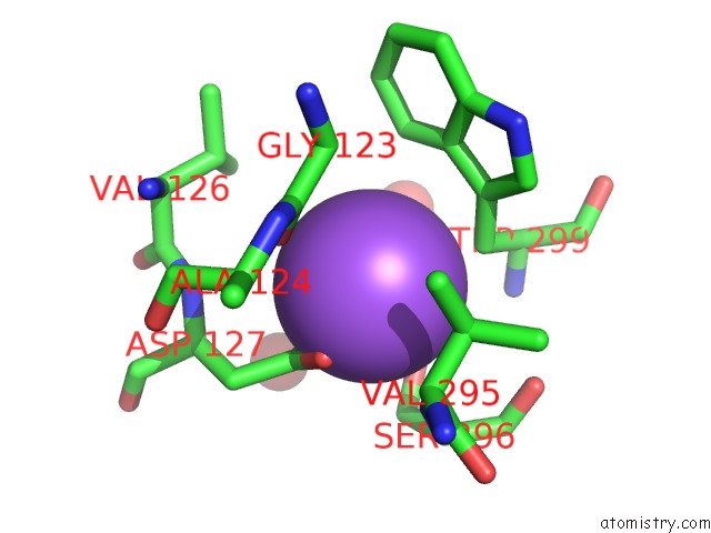
Mono view
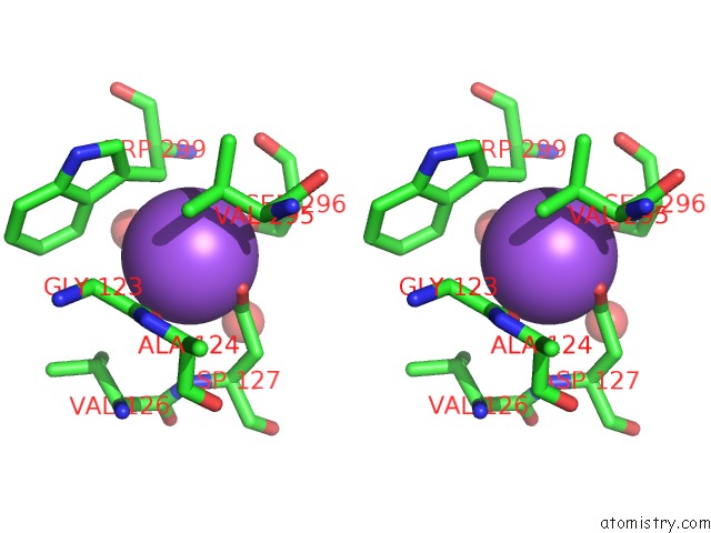
Stereo pair view
Mono view
Stereo pair view
A full contact list of Sodium with other atoms in the Na binding
site number 1 of Crystal Structure of 6-Aminohexanoate-Dimer Hydrolase S112A/G181D/H266N Mutant with Substrate within 5.0Å range:
|
Reference:
Y.Kawashima,
K.Yasuhira,
N.Shibata,
Y.Matsuura,
Y.Tanaka,
M.Taniguchi,
Y.Miyoshi,
M.Takeo,
D.Kato,
Y.Higuchi,
S.Negoro.
Enzymatic Synthesis of Nylon-6 Units in Organic Sol Contained Low-Water: Structural Requirement of 6-Aminohexanoate-Dimer Hydrolase For Efficient Amid Synthesis To Be Published.
Page generated: Mon Oct 7 05:44:25 2024
Last articles
Ca in 5NEUCa in 5NET
Ca in 5NER
Ca in 5NEM
Ca in 5NE5
Ca in 5NBP
Ca in 5NBN
Ca in 5NBM
Ca in 5NBL
Ca in 5N7G