Sodium »
PDB 2woi-2x2v »
2x0i »
Sodium in PDB 2x0i: 2.9 A Resolution Structure of Malate Dehydrogenase From Archaeoglobus Fulgidus in Complex with Nadh
Enzymatic activity of 2.9 A Resolution Structure of Malate Dehydrogenase From Archaeoglobus Fulgidus in Complex with Nadh
All present enzymatic activity of 2.9 A Resolution Structure of Malate Dehydrogenase From Archaeoglobus Fulgidus in Complex with Nadh:
1.1.1.37;
1.1.1.37;
Protein crystallography data
The structure of 2.9 A Resolution Structure of Malate Dehydrogenase From Archaeoglobus Fulgidus in Complex with Nadh, PDB code: 2x0i
was solved by
A.Irimia,
D.Madern,
G.Zaccai,
F.M.D.Vellieux,
A.Karshikoff,
G.Tibbelin,
R.Ladenstein,
T.Lien,
N.-K.Birkeland,
with X-Ray Crystallography technique. A brief refinement statistics is given in the table below:
| Resolution Low / High (Å) | 56.48 / 2.91 |
| Space group | P 41 21 2 |
| Cell size a, b, c (Å), α, β, γ (°) | 112.964, 112.964, 71.294, 90.00, 90.00, 90.00 |
| R / Rfree (%) | 15.19 / 21.86 |
Sodium Binding Sites:
The binding sites of Sodium atom in the 2.9 A Resolution Structure of Malate Dehydrogenase From Archaeoglobus Fulgidus in Complex with Nadh
(pdb code 2x0i). This binding sites where shown within
5.0 Angstroms radius around Sodium atom.
In total only one binding site of Sodium was determined in the 2.9 A Resolution Structure of Malate Dehydrogenase From Archaeoglobus Fulgidus in Complex with Nadh, PDB code: 2x0i:
In total only one binding site of Sodium was determined in the 2.9 A Resolution Structure of Malate Dehydrogenase From Archaeoglobus Fulgidus in Complex with Nadh, PDB code: 2x0i:
Sodium binding site 1 out of 1 in 2x0i
Go back to
Sodium binding site 1 out
of 1 in the 2.9 A Resolution Structure of Malate Dehydrogenase From Archaeoglobus Fulgidus in Complex with Nadh
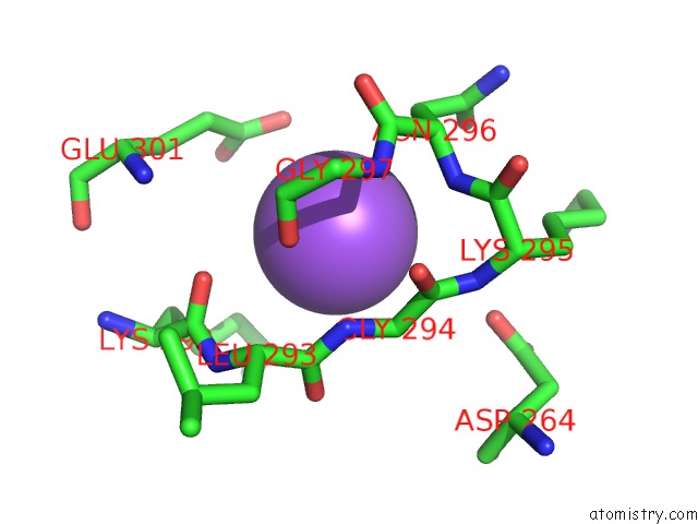
Mono view
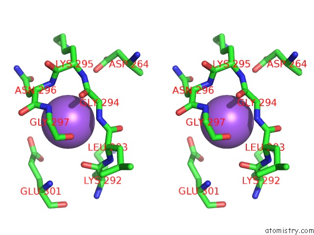
Stereo pair view
Mono view
Stereo pair view
A full contact list of Sodium with other atoms in the Na binding
site number 1 of 2.9 A Resolution Structure of Malate Dehydrogenase From Archaeoglobus Fulgidus in Complex with Nadh within 5.0Å range:
|
Reference:
A.Irimia,
F.M.D.Vellieux,
D.Madern,
G.Zaccai,
A.Karshikoff,
G.Tibbelin,
R.Ladenstein,
T.Lien,
N.-K.Birkeland.
The 2.9A Resolution Crystal Structure of Malate Dehydrogenase From Archaeoglobus Fulgidus: Mechanisms of Oligomerisation and Thermal Stabilisation. J.Mol.Biol. V. 335 343 2004.
ISSN: ISSN 0022-2836
PubMed: 14659762
DOI: 10.1016/J.JMB.2003.10.054
Page generated: Mon Oct 7 05:02:40 2024
ISSN: ISSN 0022-2836
PubMed: 14659762
DOI: 10.1016/J.JMB.2003.10.054
Last articles
Cl in 5G54Cl in 5G4A
Cl in 5G4Q
Cl in 5G47
Cl in 5G42
Cl in 5G3S
Cl in 5G2P
Cl in 5G2T
Cl in 5G36
Cl in 5G2D