Sodium »
PDB 2woi-2x2v »
2wp9 »
Sodium in PDB 2wp9: Crystal Structure of the E. Coli Succinate:Quinone Oxidoreductase (Sqr) Sdhb HIS207THR Mutant
Enzymatic activity of Crystal Structure of the E. Coli Succinate:Quinone Oxidoreductase (Sqr) Sdhb HIS207THR Mutant
All present enzymatic activity of Crystal Structure of the E. Coli Succinate:Quinone Oxidoreductase (Sqr) Sdhb HIS207THR Mutant:
1.3.5.1; 1.3.99.1;
1.3.5.1; 1.3.99.1;
Protein crystallography data
The structure of Crystal Structure of the E. Coli Succinate:Quinone Oxidoreductase (Sqr) Sdhb HIS207THR Mutant, PDB code: 2wp9
was solved by
J.Ruprecht,
V.Yankovskaya,
E.Maklashina,
S.Iwata,
G.Cecchini,
with X-Ray Crystallography technique. A brief refinement statistics is given in the table below:
| Resolution Low / High (Å) | 48.85 / 2.70 |
| Space group | P 21 21 21 |
| Cell size a, b, c (Å), α, β, γ (°) | 119.853, 183.803, 202.777, 90.00, 90.00, 90.00 |
| R / Rfree (%) | 19 / 22.2 |
Other elements in 2wp9:
The structure of Crystal Structure of the E. Coli Succinate:Quinone Oxidoreductase (Sqr) Sdhb HIS207THR Mutant also contains other interesting chemical elements:
| Iron | (Fe) | 30 atoms |
Sodium Binding Sites:
The binding sites of Sodium atom in the Crystal Structure of the E. Coli Succinate:Quinone Oxidoreductase (Sqr) Sdhb HIS207THR Mutant
(pdb code 2wp9). This binding sites where shown within
5.0 Angstroms radius around Sodium atom.
In total 3 binding sites of Sodium where determined in the Crystal Structure of the E. Coli Succinate:Quinone Oxidoreductase (Sqr) Sdhb HIS207THR Mutant, PDB code: 2wp9:
Jump to Sodium binding site number: 1; 2; 3;
In total 3 binding sites of Sodium where determined in the Crystal Structure of the E. Coli Succinate:Quinone Oxidoreductase (Sqr) Sdhb HIS207THR Mutant, PDB code: 2wp9:
Jump to Sodium binding site number: 1; 2; 3;
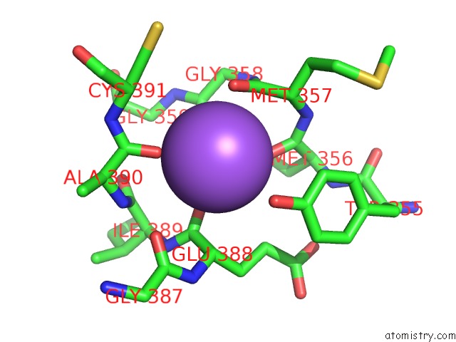
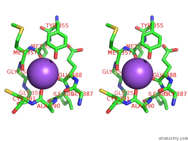
Sodium binding site 1 out of 3 in 2wp9
Go back to
Sodium binding site 1 out
of 3 in the Crystal Structure of the E. Coli Succinate:Quinone Oxidoreductase (Sqr) Sdhb HIS207THR Mutant
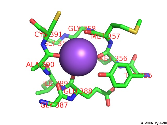
Mono view
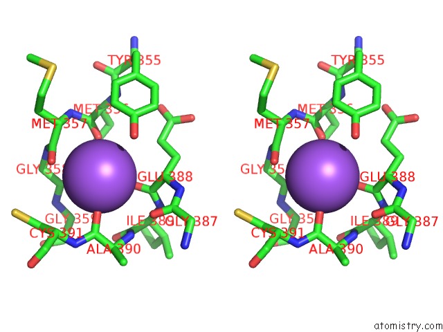
Stereo pair view
Mono view
Stereo pair view
A full contact list of Sodium with other atoms in the Na binding
site number 1 of Crystal Structure of the E. Coli Succinate:Quinone Oxidoreductase (Sqr) Sdhb HIS207THR Mutant within 5.0Å range:
|
Sodium binding site 2 out of 3 in 2wp9
Go back to
Sodium binding site 2 out
of 3 in the Crystal Structure of the E. Coli Succinate:Quinone Oxidoreductase (Sqr) Sdhb HIS207THR Mutant
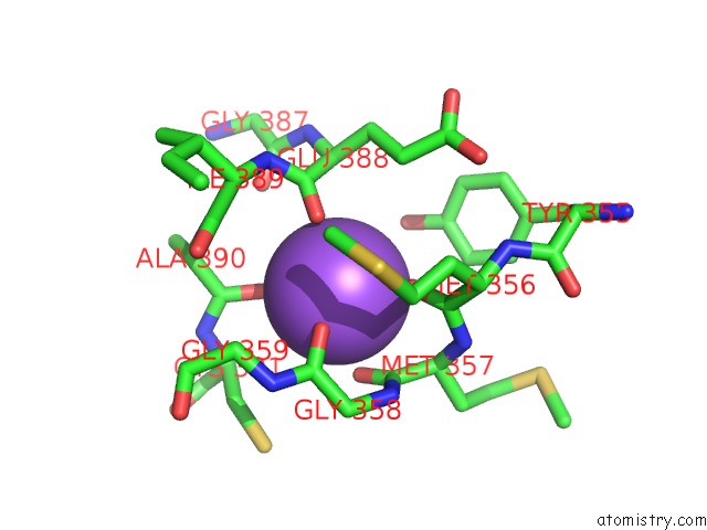
Mono view
Stereo pair view
Mono view
Stereo pair view
A full contact list of Sodium with other atoms in the Na binding
site number 2 of Crystal Structure of the E. Coli Succinate:Quinone Oxidoreductase (Sqr) Sdhb HIS207THR Mutant within 5.0Å range:
|
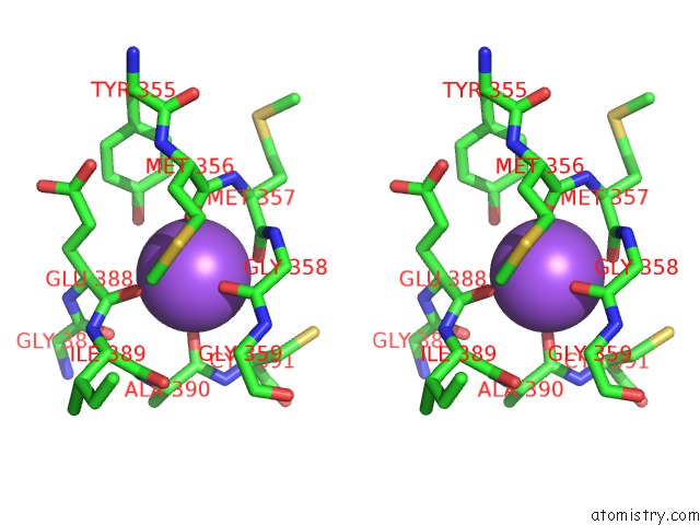
Sodium binding site 3 out of 3 in 2wp9
Go back to
Sodium binding site 3 out
of 3 in the Crystal Structure of the E. Coli Succinate:Quinone Oxidoreductase (Sqr) Sdhb HIS207THR Mutant
Mono view
Stereo pair view
Mono view
Stereo pair view
A full contact list of Sodium with other atoms in the Na binding
site number 3 of Crystal Structure of the E. Coli Succinate:Quinone Oxidoreductase (Sqr) Sdhb HIS207THR Mutant within 5.0Å range:
|
Reference:
J.Ruprecht,
S.Iwata,
R.A.Rothery,
J.H.Weiner,
E.Maklashina,
G.Cecchini.
Perturbation of the Quinone-Binding Site of Complex II Alters the Electronic Properties of the Proximal [3FE-4S] Iron-Sulfur Cluster. J. Biol. Chem. V. 286 12756 2011.
ISSN: ESSN 1083-351X
PubMed: 21310949
DOI: 10.1074/JBC.M110.209874
Page generated: Mon Oct 7 04:51:37 2024
ISSN: ESSN 1083-351X
PubMed: 21310949
DOI: 10.1074/JBC.M110.209874
Last articles
Fe in 2YXOFe in 2YRS
Fe in 2YXC
Fe in 2YNM
Fe in 2YVJ
Fe in 2YP1
Fe in 2YU2
Fe in 2YU1
Fe in 2YQB
Fe in 2YOO