Sodium »
PDB 2uzz-2vrp »
2v4v »
Sodium in PDB 2v4v: Crystal Structure of A Family 6 Carbohydrate-Binding Module From Clostridium Cellulolyticum in Complex with Xylose
Protein crystallography data
The structure of Crystal Structure of A Family 6 Carbohydrate-Binding Module From Clostridium Cellulolyticum in Complex with Xylose, PDB code: 2v4v
was solved by
D.W.Abbott,
E.Ficko-Blean,
A.Lammerts Van Bueren,
P.M.Coutinho,
B.Henrissat,
H.J.Gilbert,
A.B.Boraston,
with X-Ray Crystallography technique. A brief refinement statistics is given in the table below:
| Resolution Low / High (Å) | 93.25 / 1.50 |
| Space group | P 21 21 21 |
| Cell size a, b, c (Å), α, β, γ (°) | 31.026, 43.598, 93.323, 90.00, 90.00, 90.00 |
| R / Rfree (%) | 19 / 22.2 |
Sodium Binding Sites:
The binding sites of Sodium atom in the Crystal Structure of A Family 6 Carbohydrate-Binding Module From Clostridium Cellulolyticum in Complex with Xylose
(pdb code 2v4v). This binding sites where shown within
5.0 Angstroms radius around Sodium atom.
In total only one binding site of Sodium was determined in the Crystal Structure of A Family 6 Carbohydrate-Binding Module From Clostridium Cellulolyticum in Complex with Xylose, PDB code: 2v4v:
In total only one binding site of Sodium was determined in the Crystal Structure of A Family 6 Carbohydrate-Binding Module From Clostridium Cellulolyticum in Complex with Xylose, PDB code: 2v4v:
Sodium binding site 1 out of 1 in 2v4v
Go back to
Sodium binding site 1 out
of 1 in the Crystal Structure of A Family 6 Carbohydrate-Binding Module From Clostridium Cellulolyticum in Complex with Xylose
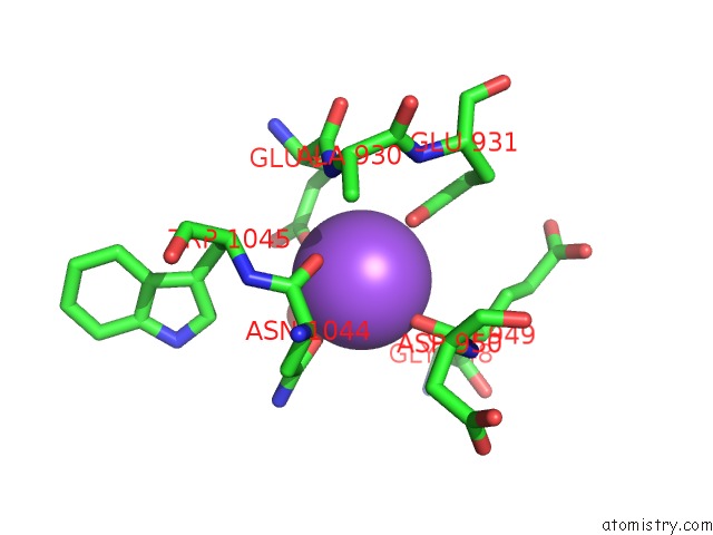
Mono view
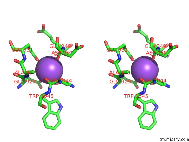
Stereo pair view
Mono view
Stereo pair view
A full contact list of Sodium with other atoms in the Na binding
site number 1 of Crystal Structure of A Family 6 Carbohydrate-Binding Module From Clostridium Cellulolyticum in Complex with Xylose within 5.0Å range:
|
Reference:
D.W.Abbott,
E.Ficko-Blean,
A.L.Van Bueren,
A.Rogowski,
A.Cartmell,
P.M.Coutinho,
B.Henrissat,
H.J.Gilbert,
A.B.Boraston.
Analysis of the Structural and Functional Diversity of Plant Cell Wall Specific Family 6 Carbohydrate Binding Modules. Biochemistry V. 48 10395 2009.
ISSN: ISSN 0006-2960
PubMed: 19788273
DOI: 10.1021/BI9013424
Page generated: Mon Oct 7 04:26:18 2024
ISSN: ISSN 0006-2960
PubMed: 19788273
DOI: 10.1021/BI9013424
Last articles
Zn in 9MJ5Zn in 9HNW
Zn in 9G0L
Zn in 9FNE
Zn in 9DZN
Zn in 9E0I
Zn in 9D32
Zn in 9DAK
Zn in 8ZXC
Zn in 8ZUF