Sodium »
PDB 2ply-2qd6 »
2pnk »
Sodium in PDB 2pnk: Crystal Structure of An Uronate Isomerase (BH0493) From Bacillus Halodurans C-125 at 2.00 A Resolution
Protein crystallography data
The structure of Crystal Structure of An Uronate Isomerase (BH0493) From Bacillus Halodurans C-125 at 2.00 A Resolution, PDB code: 2pnk
was solved by
Joint Center For Structural Genomics (Jcsg),
with X-Ray Crystallography technique. A brief refinement statistics is given in the table below:
| Resolution Low / High (Å) | 48.56 / 2.00 |
| Space group | C 1 2 1 |
| Cell size a, b, c (Å), α, β, γ (°) | 273.720, 158.560, 181.240, 90.00, 116.03, 90.00 |
| R / Rfree (%) | 14.8 / 17.8 |
Other elements in 2pnk:
The structure of Crystal Structure of An Uronate Isomerase (BH0493) From Bacillus Halodurans C-125 at 2.00 A Resolution also contains other interesting chemical elements:
| Arsenic | (As) | 3 atoms |
| Chlorine | (Cl) | 8 atoms |
Sodium Binding Sites:
The binding sites of Sodium atom in the Crystal Structure of An Uronate Isomerase (BH0493) From Bacillus Halodurans C-125 at 2.00 A Resolution
(pdb code 2pnk). This binding sites where shown within
5.0 Angstroms radius around Sodium atom.
In total 3 binding sites of Sodium where determined in the Crystal Structure of An Uronate Isomerase (BH0493) From Bacillus Halodurans C-125 at 2.00 A Resolution, PDB code: 2pnk:
Jump to Sodium binding site number: 1; 2; 3;
In total 3 binding sites of Sodium where determined in the Crystal Structure of An Uronate Isomerase (BH0493) From Bacillus Halodurans C-125 at 2.00 A Resolution, PDB code: 2pnk:
Jump to Sodium binding site number: 1; 2; 3;
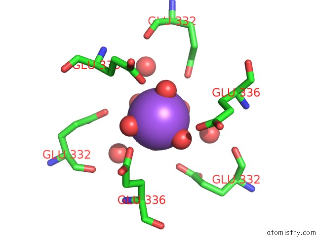
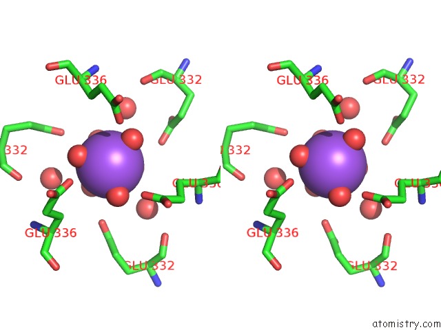
Sodium binding site 1 out of 3 in 2pnk
Go back to
Sodium binding site 1 out
of 3 in the Crystal Structure of An Uronate Isomerase (BH0493) From Bacillus Halodurans C-125 at 2.00 A Resolution
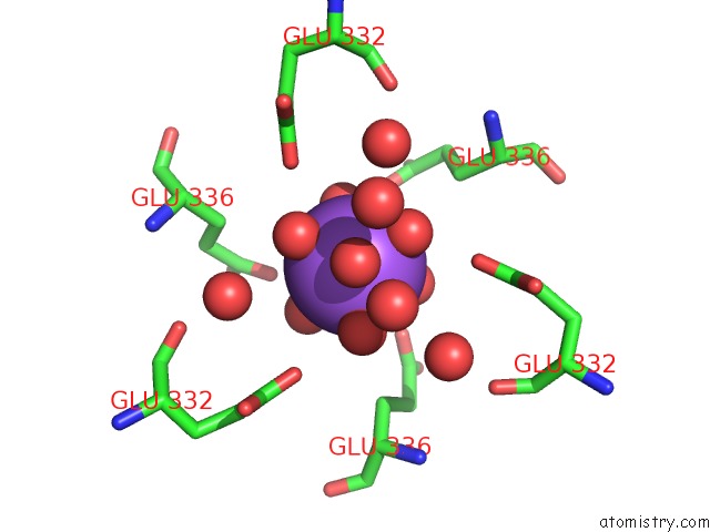
Mono view
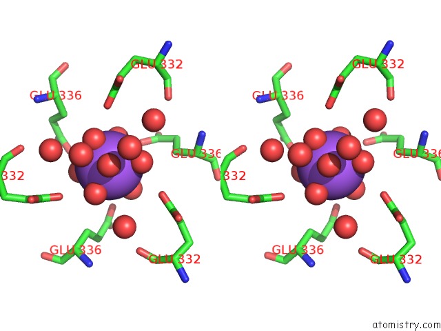
Stereo pair view
Mono view
Stereo pair view
A full contact list of Sodium with other atoms in the Na binding
site number 1 of Crystal Structure of An Uronate Isomerase (BH0493) From Bacillus Halodurans C-125 at 2.00 A Resolution within 5.0Å range:
|
Sodium binding site 2 out of 3 in 2pnk
Go back to
Sodium binding site 2 out
of 3 in the Crystal Structure of An Uronate Isomerase (BH0493) From Bacillus Halodurans C-125 at 2.00 A Resolution
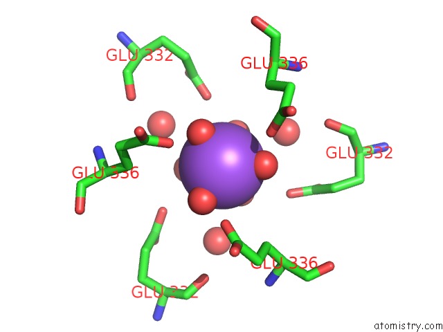
Mono view
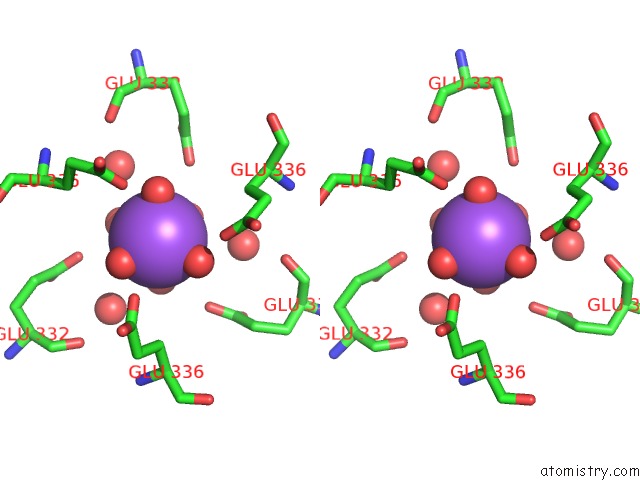
Stereo pair view
Mono view
Stereo pair view
A full contact list of Sodium with other atoms in the Na binding
site number 2 of Crystal Structure of An Uronate Isomerase (BH0493) From Bacillus Halodurans C-125 at 2.00 A Resolution within 5.0Å range:
|
Sodium binding site 3 out of 3 in 2pnk
Go back to
Sodium binding site 3 out
of 3 in the Crystal Structure of An Uronate Isomerase (BH0493) From Bacillus Halodurans C-125 at 2.00 A Resolution
Mono view
Stereo pair view
Mono view
Stereo pair view
A full contact list of Sodium with other atoms in the Na binding
site number 3 of Crystal Structure of An Uronate Isomerase (BH0493) From Bacillus Halodurans C-125 at 2.00 A Resolution within 5.0Å range:
|
Reference:
Joint Center For Structural Genomics (Jcsg),
Joint Center For Structural Genomics (Jcsg).
N/A N/A.
Page generated: Sun Aug 17 11:21:12 2025
Last articles
Na in 4J20Na in 4J1H
Na in 4J1F
Na in 4J1E
Na in 4J1C
Na in 4J17
Na in 4J0Z
Na in 4J1B
Na in 4J1A
Na in 4J0T