Sodium »
PDB 2isz-2jln »
2jbw »
Sodium in PDB 2jbw: Crystal Structure of the 2,6-Dihydroxy-Pseudo-Oxynicotine Hydrolase.
Protein crystallography data
The structure of Crystal Structure of the 2,6-Dihydroxy-Pseudo-Oxynicotine Hydrolase., PDB code: 2jbw
was solved by
C.Schleberger,
P.Sachelaru,
R.Brandsch,
G.E.Schulz,
with X-Ray Crystallography technique. A brief refinement statistics is given in the table below:
| Resolution Low / High (Å) | 30.00 / 2.10 |
| Space group | P 1 21 1 |
| Cell size a, b, c (Å), α, β, γ (°) | 90.570, 57.020, 152.697, 90.00, 103.35, 90.00 |
| R / Rfree (%) | 18.1 / 23.8 |
Sodium Binding Sites:
The binding sites of Sodium atom in the Crystal Structure of the 2,6-Dihydroxy-Pseudo-Oxynicotine Hydrolase.
(pdb code 2jbw). This binding sites where shown within
5.0 Angstroms radius around Sodium atom.
In total 4 binding sites of Sodium where determined in the Crystal Structure of the 2,6-Dihydroxy-Pseudo-Oxynicotine Hydrolase., PDB code: 2jbw:
Jump to Sodium binding site number: 1; 2; 3; 4;
In total 4 binding sites of Sodium where determined in the Crystal Structure of the 2,6-Dihydroxy-Pseudo-Oxynicotine Hydrolase., PDB code: 2jbw:
Jump to Sodium binding site number: 1; 2; 3; 4;
Sodium binding site 1 out of 4 in 2jbw
Go back to
Sodium binding site 1 out
of 4 in the Crystal Structure of the 2,6-Dihydroxy-Pseudo-Oxynicotine Hydrolase.
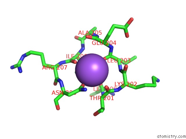
Mono view

Stereo pair view
Mono view
Stereo pair view
A full contact list of Sodium with other atoms in the Na binding
site number 1 of Crystal Structure of the 2,6-Dihydroxy-Pseudo-Oxynicotine Hydrolase. within 5.0Å range:
|
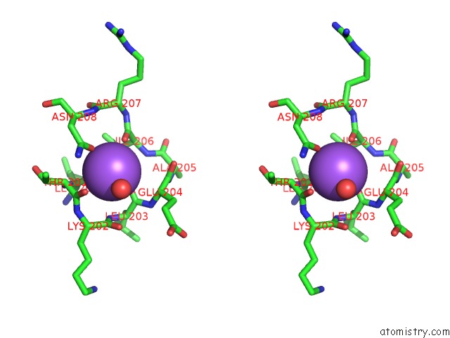
Sodium binding site 2 out of 4 in 2jbw
Go back to
Sodium binding site 2 out
of 4 in the Crystal Structure of the 2,6-Dihydroxy-Pseudo-Oxynicotine Hydrolase.

Mono view
Stereo pair view
Mono view
Stereo pair view
A full contact list of Sodium with other atoms in the Na binding
site number 2 of Crystal Structure of the 2,6-Dihydroxy-Pseudo-Oxynicotine Hydrolase. within 5.0Å range:
|
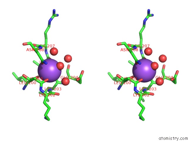
Sodium binding site 3 out of 4 in 2jbw
Go back to
Sodium binding site 3 out
of 4 in the Crystal Structure of the 2,6-Dihydroxy-Pseudo-Oxynicotine Hydrolase.

Mono view
Stereo pair view
Mono view
Stereo pair view
A full contact list of Sodium with other atoms in the Na binding
site number 3 of Crystal Structure of the 2,6-Dihydroxy-Pseudo-Oxynicotine Hydrolase. within 5.0Å range:
|
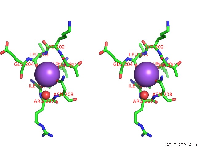
Sodium binding site 4 out of 4 in 2jbw
Go back to
Sodium binding site 4 out
of 4 in the Crystal Structure of the 2,6-Dihydroxy-Pseudo-Oxynicotine Hydrolase.

Mono view
Stereo pair view
Mono view
Stereo pair view
A full contact list of Sodium with other atoms in the Na binding
site number 4 of Crystal Structure of the 2,6-Dihydroxy-Pseudo-Oxynicotine Hydrolase. within 5.0Å range:
|
Reference:
C.Schleberger,
P.Sachelaru,
R.Brandsch,
G.E.Schulz.
Structure and Action of A Cc Bond Cleaving Alpha/Beta-Hydrolase Involved in Nicotine Degration. J.Mol.Biol. V. 367 409 2007.
ISSN: ISSN 0022-2836
PubMed: 17275835
DOI: 10.1016/J.JMB.2006.12.068
Page generated: Mon Oct 7 02:59:56 2024
ISSN: ISSN 0022-2836
PubMed: 17275835
DOI: 10.1016/J.JMB.2006.12.068
Last articles
Zn in 9J0NZn in 9J0O
Zn in 9J0P
Zn in 9FJX
Zn in 9EKB
Zn in 9C0F
Zn in 9CAH
Zn in 9CH0
Zn in 9CH3
Zn in 9CH1