Sodium »
PDB 2htx-2isp »
2iaj »
Sodium in PDB 2iaj: Crystal Structure of K103N/Y181C Mutant Hiv-1 Reverse Transcriptase (Rt) in Complex with Atp
Enzymatic activity of Crystal Structure of K103N/Y181C Mutant Hiv-1 Reverse Transcriptase (Rt) in Complex with Atp
All present enzymatic activity of Crystal Structure of K103N/Y181C Mutant Hiv-1 Reverse Transcriptase (Rt) in Complex with Atp:
2.7.7.49;
2.7.7.49;
Protein crystallography data
The structure of Crystal Structure of K103N/Y181C Mutant Hiv-1 Reverse Transcriptase (Rt) in Complex with Atp, PDB code: 2iaj
was solved by
K.Das,
E.Arnold,
with X-Ray Crystallography technique. A brief refinement statistics is given in the table below:
| Resolution Low / High (Å) | 19.96 / 2.50 |
| Space group | C 1 2 1 |
| Cell size a, b, c (Å), α, β, γ (°) | 237.400, 71.130, 94.210, 90.00, 105.86, 90.00 |
| R / Rfree (%) | 23.3 / 28.4 |
Other elements in 2iaj:
The structure of Crystal Structure of K103N/Y181C Mutant Hiv-1 Reverse Transcriptase (Rt) in Complex with Atp also contains other interesting chemical elements:
| Manganese | (Mn) | 3 atoms |
Sodium Binding Sites:
The binding sites of Sodium atom in the Crystal Structure of K103N/Y181C Mutant Hiv-1 Reverse Transcriptase (Rt) in Complex with Atp
(pdb code 2iaj). This binding sites where shown within
5.0 Angstroms radius around Sodium atom.
In total only one binding site of Sodium was determined in the Crystal Structure of K103N/Y181C Mutant Hiv-1 Reverse Transcriptase (Rt) in Complex with Atp, PDB code: 2iaj:
In total only one binding site of Sodium was determined in the Crystal Structure of K103N/Y181C Mutant Hiv-1 Reverse Transcriptase (Rt) in Complex with Atp, PDB code: 2iaj:
Sodium binding site 1 out of 1 in 2iaj
Go back to
Sodium binding site 1 out
of 1 in the Crystal Structure of K103N/Y181C Mutant Hiv-1 Reverse Transcriptase (Rt) in Complex with Atp
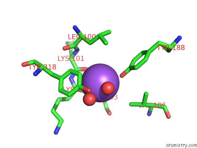
Mono view
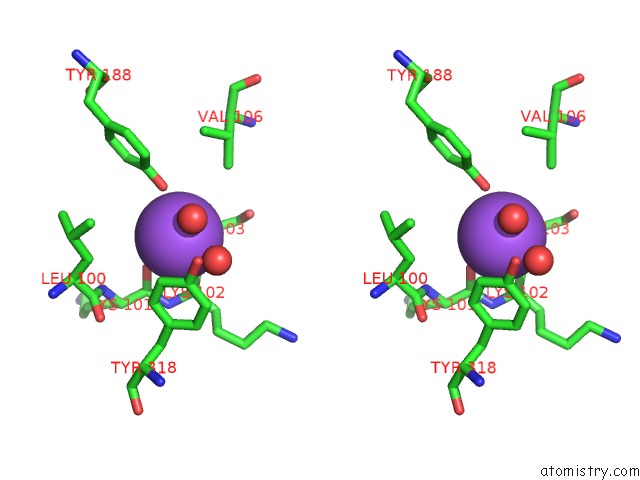
Stereo pair view
Mono view
Stereo pair view
A full contact list of Sodium with other atoms in the Na binding
site number 1 of Crystal Structure of K103N/Y181C Mutant Hiv-1 Reverse Transcriptase (Rt) in Complex with Atp within 5.0Å range:
|
Reference:
K.Das,
S.G.Sarafianos,
A.D.Clark,
P.L.Boyer,
S.H.Hughes,
E.Arnold.
Crystal Structures of Clinically Relevant LYS103ASN/TYR181CYS Double Mutant Hiv-1 Reverse Transcriptase in Complexes with Atp and Non-Nucleoside Inhibitor Hby 097. J.Mol.Biol. V. 365 77 2007.
ISSN: ISSN 0022-2836
PubMed: 17056061
DOI: 10.1016/J.JMB.2006.08.097
Page generated: Mon Oct 7 02:51:34 2024
ISSN: ISSN 0022-2836
PubMed: 17056061
DOI: 10.1016/J.JMB.2006.08.097
Last articles
Cl in 5TPICl in 5TQI
Cl in 5TPU
Cl in 5TPH
Cl in 5TPX
Cl in 5TPG
Cl in 5TOW
Cl in 5TNU
Cl in 5TPA
Cl in 5TOV