Sodium »
PDB 2gum-2hti »
2hrw »
Sodium in PDB 2hrw: Crystal Structure of Phosphonopyruvate Hydrolase
Protein crystallography data
The structure of Crystal Structure of Phosphonopyruvate Hydrolase, PDB code: 2hrw
was solved by
C.C.H.Chen,
O.Herzberg,
with X-Ray Crystallography technique. A brief refinement statistics is given in the table below:
| Resolution Low / High (Å) | 32.62 / 2.20 |
| Space group | I 2 2 2 |
| Cell size a, b, c (Å), α, β, γ (°) | 74.880, 79.020, 93.440, 90.00, 90.00, 90.00 |
| R / Rfree (%) | 20.4 / 27.1 |
Other elements in 2hrw:
The structure of Crystal Structure of Phosphonopyruvate Hydrolase also contains other interesting chemical elements:
| Chlorine | (Cl) | 2 atoms |
Sodium Binding Sites:
The binding sites of Sodium atom in the Crystal Structure of Phosphonopyruvate Hydrolase
(pdb code 2hrw). This binding sites where shown within
5.0 Angstroms radius around Sodium atom.
In total only one binding site of Sodium was determined in the Crystal Structure of Phosphonopyruvate Hydrolase, PDB code: 2hrw:
In total only one binding site of Sodium was determined in the Crystal Structure of Phosphonopyruvate Hydrolase, PDB code: 2hrw:
Sodium binding site 1 out of 1 in 2hrw
Go back to
Sodium binding site 1 out
of 1 in the Crystal Structure of Phosphonopyruvate Hydrolase
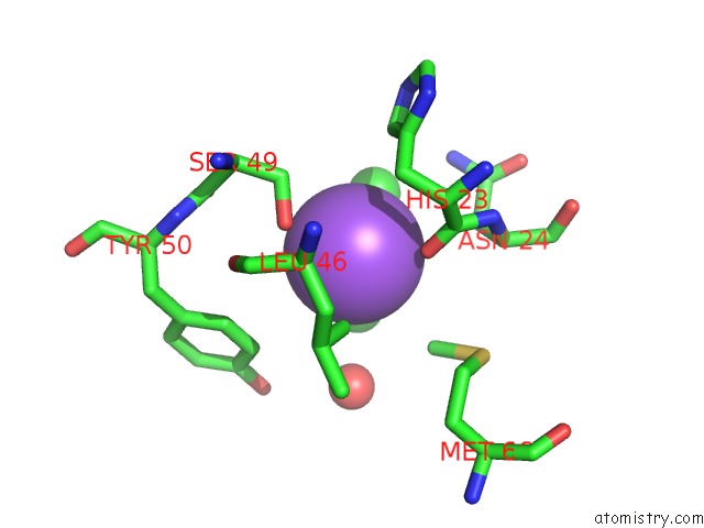
Mono view
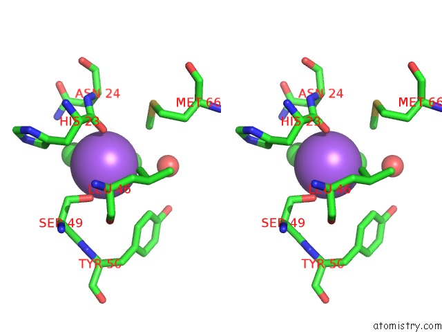
Stereo pair view
Mono view
Stereo pair view
A full contact list of Sodium with other atoms in the Na binding
site number 1 of Crystal Structure of Phosphonopyruvate Hydrolase within 5.0Å range:
|
Reference:
C.C.H.Chen,
Y.Han,
W.Niu,
A.N.Kulakova,
A.Howard,
J.P.Quinn,
D.Dunaway-Mariano,
O.Herzberg.
Structure and Kinetics of Phosphonopyruvate Hydrolase From Voriovorax Sp. PAL2: New Insight Into the Divergence of Catalysis Within the Pep Mutase/Isocitrate Lyase Superfamily Biochemistry V. 45 11491 2006.
ISSN: ISSN 0006-2960
PubMed: 16981709
DOI: 10.1021/BI061208L
Page generated: Mon Oct 7 02:47:55 2024
ISSN: ISSN 0006-2960
PubMed: 16981709
DOI: 10.1021/BI061208L
Last articles
Zn in 9MJ5Zn in 9HNW
Zn in 9G0L
Zn in 9FNE
Zn in 9DZN
Zn in 9E0I
Zn in 9D32
Zn in 9DAK
Zn in 8ZXC
Zn in 8ZUF