Sodium »
PDB 2gum-2hti »
2gxb »
Sodium in PDB 2gxb: Crystal Structure of the Za Domain Bound to Z-Rna
Protein crystallography data
The structure of Crystal Structure of the Za Domain Bound to Z-Rna, PDB code: 2gxb
was solved by
A.Athanasiadis,
D.Placido,
A.Rich,
with X-Ray Crystallography technique. A brief refinement statistics is given in the table below:
| Resolution Low / High (Å) | 25.00 / 2.25 |
| Space group | C 2 2 21 |
| Cell size a, b, c (Å), α, β, γ (°) | 73.603, 92.776, 50.229, 90.00, 90.00, 90.00 |
| R / Rfree (%) | 19.4 / 24.4 |
Sodium Binding Sites:
The binding sites of Sodium atom in the Crystal Structure of the Za Domain Bound to Z-Rna
(pdb code 2gxb). This binding sites where shown within
5.0 Angstroms radius around Sodium atom.
In total 3 binding sites of Sodium where determined in the Crystal Structure of the Za Domain Bound to Z-Rna, PDB code: 2gxb:
Jump to Sodium binding site number: 1; 2; 3;
In total 3 binding sites of Sodium where determined in the Crystal Structure of the Za Domain Bound to Z-Rna, PDB code: 2gxb:
Jump to Sodium binding site number: 1; 2; 3;
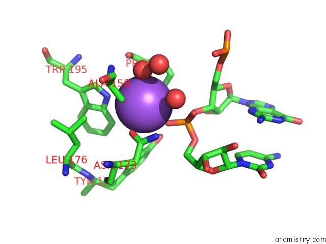
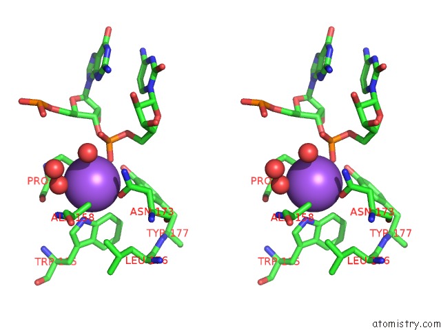
Sodium binding site 1 out of 3 in 2gxb
Go back to
Sodium binding site 1 out
of 3 in the Crystal Structure of the Za Domain Bound to Z-Rna
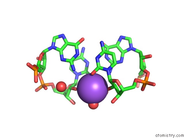
Mono view
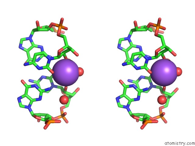
Stereo pair view
Mono view
Stereo pair view
A full contact list of Sodium with other atoms in the Na binding
site number 1 of Crystal Structure of the Za Domain Bound to Z-Rna within 5.0Å range:
|
Sodium binding site 2 out of 3 in 2gxb
Go back to
Sodium binding site 2 out
of 3 in the Crystal Structure of the Za Domain Bound to Z-Rna
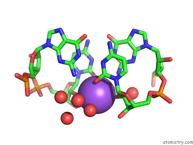
Mono view
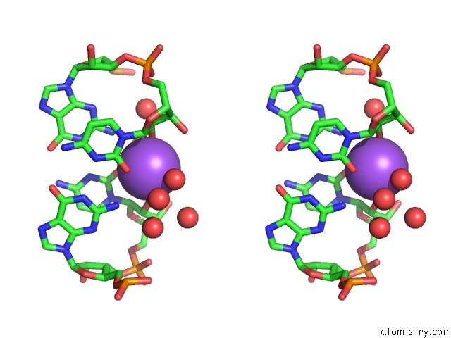
Stereo pair view
Mono view
Stereo pair view
A full contact list of Sodium with other atoms in the Na binding
site number 2 of Crystal Structure of the Za Domain Bound to Z-Rna within 5.0Å range:
|
Sodium binding site 3 out of 3 in 2gxb
Go back to
Sodium binding site 3 out
of 3 in the Crystal Structure of the Za Domain Bound to Z-Rna
Mono view
Stereo pair view
Mono view
Stereo pair view
A full contact list of Sodium with other atoms in the Na binding
site number 3 of Crystal Structure of the Za Domain Bound to Z-Rna within 5.0Å range:
|
Reference:
D.Placido,
B.A.Brown,
K.Lowenhaupt,
A.Rich,
A.Athanasiadis.
A Left-Handed Rna Double Helix Bound By the Zalpha Domain of the Rna-Editing Enzyme ADAR1. Structure V. 15 395 2007.
ISSN: ISSN 0969-2126
PubMed: 17437712
DOI: 10.1016/J.STR.2007.03.001
Page generated: Mon Oct 7 02:43:11 2024
ISSN: ISSN 0969-2126
PubMed: 17437712
DOI: 10.1016/J.STR.2007.03.001
Last articles
Zn in 9J0NZn in 9J0O
Zn in 9J0P
Zn in 9FJX
Zn in 9EKB
Zn in 9C0F
Zn in 9CAH
Zn in 9CH0
Zn in 9CH3
Zn in 9CH1