Sodium »
PDB 2gum-2hti »
2gvk »
Sodium in PDB 2gvk: Crystal Structure of A Dye-Decolorizing Peroxidase (Dyp) From Bacteroides Thetaiotaomicron Vpi-5482 at 1.6 A Resolution
Protein crystallography data
The structure of Crystal Structure of A Dye-Decolorizing Peroxidase (Dyp) From Bacteroides Thetaiotaomicron Vpi-5482 at 1.6 A Resolution, PDB code: 2gvk
was solved by
Joint Center For Structural Genomics (Jcsg),
with X-Ray Crystallography technique. A brief refinement statistics is given in the table below:
| Resolution Low / High (Å) | 28.01 / 1.60 |
| Space group | P 63 2 2 |
| Cell size a, b, c (Å), α, β, γ (°) | 109.090, 109.090, 122.100, 90.00, 90.00, 120.00 |
| R / Rfree (%) | 16.4 / 19 |
Other elements in 2gvk:
The structure of Crystal Structure of A Dye-Decolorizing Peroxidase (Dyp) From Bacteroides Thetaiotaomicron Vpi-5482 at 1.6 A Resolution also contains other interesting chemical elements:
| Chlorine | (Cl) | 3 atoms |
Sodium Binding Sites:
The binding sites of Sodium atom in the Crystal Structure of A Dye-Decolorizing Peroxidase (Dyp) From Bacteroides Thetaiotaomicron Vpi-5482 at 1.6 A Resolution
(pdb code 2gvk). This binding sites where shown within
5.0 Angstroms radius around Sodium atom.
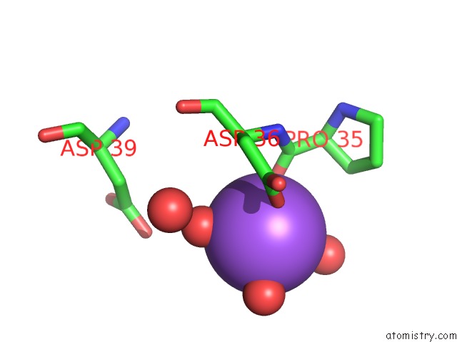
In total only one binding site of Sodium was determined in the Crystal Structure of A Dye-Decolorizing Peroxidase (Dyp) From Bacteroides Thetaiotaomicron Vpi-5482 at 1.6 A Resolution, PDB code: 2gvk:
In total only one binding site of Sodium was determined in the Crystal Structure of A Dye-Decolorizing Peroxidase (Dyp) From Bacteroides Thetaiotaomicron Vpi-5482 at 1.6 A Resolution, PDB code: 2gvk:
Sodium binding site 1 out of 1 in 2gvk
Go back to
Sodium binding site 1 out
of 1 in the Crystal Structure of A Dye-Decolorizing Peroxidase (Dyp) From Bacteroides Thetaiotaomicron Vpi-5482 at 1.6 A Resolution
Mono view

Stereo pair view
Mono view
Stereo pair view
A full contact list of Sodium with other atoms in the Na binding
site number 1 of Crystal Structure of A Dye-Decolorizing Peroxidase (Dyp) From Bacteroides Thetaiotaomicron Vpi-5482 at 1.6 A Resolution within 5.0Å range:
|
Reference:
C.Zubieta,
S.S.Krishna,
M.Kapoor,
P.Kozbial,
D.Mcmullan,
H.L.Axelrod,
M.D.Miller,
P.Abdubek,
E.Ambing,
T.Astakhova,
D.Carlton,
H.J.Chiu,
T.Clayton,
M.C.Deller,
L.Duan,
M.A.Elsliger,
J.Feuerhelm,
S.K.Grzechnik,
J.Hale,
E.Hampton,
G.W.Han,
L.Jaroszewski,
K.K.Jin,
H.E.Klock,
M.W.Knuth,
A.Kumar,
D.Marciano,
A.T.Morse,
E.Nigoghossian,
L.Okach,
S.Oommachen,
R.Reyes,
C.L.Rife,
P.Schimmel,
H.Van Den Bedem,
D.Weekes,
A.White,
Q.Xu,
K.O.Hodgson,
J.Wooley,
A.M.Deacon,
A.Godzik,
S.A.Lesley,
I.A.Wilson.
Crystal Structures of Two Novel Dye-Decolorizing Peroxidases Reveal A Beta-Barrel Fold with A Conserved Heme-Binding Motif. Proteins V. 69 223 2007.
ISSN: ISSN 0887-3585
PubMed: 17654545
DOI: 10.1002/PROT.21550
Page generated: Mon Oct 7 02:43:11 2024
ISSN: ISSN 0887-3585
PubMed: 17654545
DOI: 10.1002/PROT.21550
Last articles
Zn in 9J0NZn in 9J0O
Zn in 9J0P
Zn in 9FJX
Zn in 9EKB
Zn in 9C0F
Zn in 9CAH
Zn in 9CH0
Zn in 9CH3
Zn in 9CH1