Sodium »
PDB 2gg3-2gu7 »
2gkm »
Sodium in PDB 2gkm: Crystal Structure of Mycobacterium Tuberculosis Trhbn TYRB10PHE Mutant
Protein crystallography data
The structure of Crystal Structure of Mycobacterium Tuberculosis Trhbn TYRB10PHE Mutant, PDB code: 2gkm
was solved by
M.Milani,
M.Bolognesi,
with X-Ray Crystallography technique. A brief refinement statistics is given in the table below:
| Resolution Low / High (Å) | 51.10 / 1.73 |
| Space group | P 21 21 21 |
| Cell size a, b, c (Å), α, β, γ (°) | 45.278, 61.932, 90.291, 90.00, 90.00, 90.00 |
| R / Rfree (%) | 19.3 / 23.3 |
Other elements in 2gkm:
The structure of Crystal Structure of Mycobacterium Tuberculosis Trhbn TYRB10PHE Mutant also contains other interesting chemical elements:
| Iron | (Fe) | 2 atoms |
Sodium Binding Sites:
The binding sites of Sodium atom in the Crystal Structure of Mycobacterium Tuberculosis Trhbn TYRB10PHE Mutant
(pdb code 2gkm). This binding sites where shown within
5.0 Angstroms radius around Sodium atom.
In total only one binding site of Sodium was determined in the Crystal Structure of Mycobacterium Tuberculosis Trhbn TYRB10PHE Mutant, PDB code: 2gkm:
In total only one binding site of Sodium was determined in the Crystal Structure of Mycobacterium Tuberculosis Trhbn TYRB10PHE Mutant, PDB code: 2gkm:
Sodium binding site 1 out of 1 in 2gkm
Go back to
Sodium binding site 1 out
of 1 in the Crystal Structure of Mycobacterium Tuberculosis Trhbn TYRB10PHE Mutant

Mono view
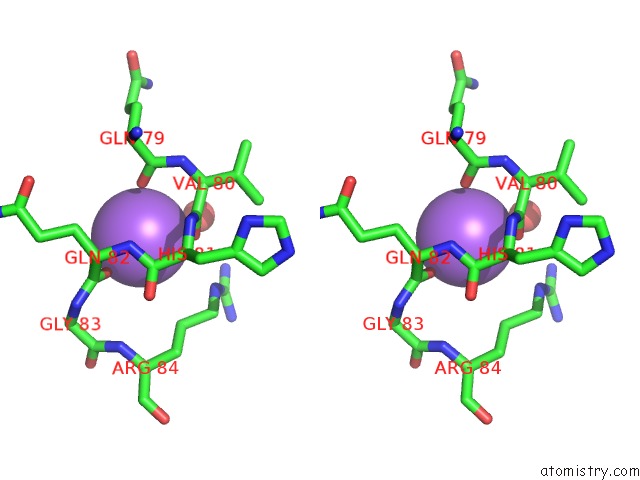
Stereo pair view
Mono view
Stereo pair view
A full contact list of Sodium with other atoms in the Na binding
site number 1 of Crystal Structure of Mycobacterium Tuberculosis Trhbn TYRB10PHE Mutant within 5.0Å range:
|
Reference:
Y.Ouellet,
M.Milani,
M.Couture,
M.Bolognesi,
M.Guertin.
Ligand Interactions in the Distal Heme Pocket of Mycobacterium Tuberculosis Truncated Hemoglobin N: Roles of TYRB10 and GLNE11 Residues Biochemistry V. 45 8770 2006.
ISSN: ISSN 0006-2960
PubMed: 16846220
DOI: 10.1021/BI060112O
Page generated: Mon Oct 7 02:39:00 2024
ISSN: ISSN 0006-2960
PubMed: 16846220
DOI: 10.1021/BI060112O
Last articles
Zn in 9J0NZn in 9J0O
Zn in 9J0P
Zn in 9FJX
Zn in 9EKB
Zn in 9C0F
Zn in 9CAH
Zn in 9CH0
Zn in 9CH3
Zn in 9CH1