Sodium »
PDB 2ekb-2fm1 »
2fm1 »
Sodium in PDB 2fm1: Crystal Structure of L-Allo-Threonine Aldolase (TM1744) From Thermotoga Maritima at 2.25 A Resolution
Protein crystallography data
The structure of Crystal Structure of L-Allo-Threonine Aldolase (TM1744) From Thermotoga Maritima at 2.25 A Resolution, PDB code: 2fm1
was solved by
Joint Center For Structural Genomics (Jcsg),
with X-Ray Crystallography technique. A brief refinement statistics is given in the table below:
| Resolution Low / High (Å) | 83.33 / 2.25 |
| Space group | P 21 21 21 |
| Cell size a, b, c (Å), α, β, γ (°) | 95.614, 100.405, 149.648, 90.00, 90.00, 90.00 |
| R / Rfree (%) | 14.2 / 21.6 |
Other elements in 2fm1:
The structure of Crystal Structure of L-Allo-Threonine Aldolase (TM1744) From Thermotoga Maritima at 2.25 A Resolution also contains other interesting chemical elements:
| Chlorine | (Cl) | 10 atoms |
| Calcium | (Ca) | 2 atoms |
Sodium Binding Sites:
The binding sites of Sodium atom in the Crystal Structure of L-Allo-Threonine Aldolase (TM1744) From Thermotoga Maritima at 2.25 A Resolution
(pdb code 2fm1). This binding sites where shown within
5.0 Angstroms radius around Sodium atom.
In total only one binding site of Sodium was determined in the Crystal Structure of L-Allo-Threonine Aldolase (TM1744) From Thermotoga Maritima at 2.25 A Resolution, PDB code: 2fm1:
In total only one binding site of Sodium was determined in the Crystal Structure of L-Allo-Threonine Aldolase (TM1744) From Thermotoga Maritima at 2.25 A Resolution, PDB code: 2fm1:
Sodium binding site 1 out of 1 in 2fm1
Go back to
Sodium binding site 1 out
of 1 in the Crystal Structure of L-Allo-Threonine Aldolase (TM1744) From Thermotoga Maritima at 2.25 A Resolution
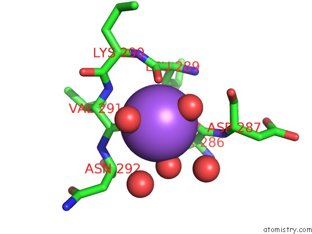
Mono view
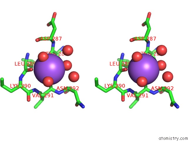
Stereo pair view
Mono view
Stereo pair view
A full contact list of Sodium with other atoms in the Na binding
site number 1 of Crystal Structure of L-Allo-Threonine Aldolase (TM1744) From Thermotoga Maritima at 2.25 A Resolution within 5.0Å range:
|
Reference:
Joint Center For Structural Genomics (Jcsg),
Joint Center For Structural Genomics (Jcsg).
N/A N/A.
Page generated: Mon Oct 7 02:27:56 2024
Last articles
Cl in 5GS8Cl in 5GQQ
Cl in 5G6U
Cl in 5GQN
Cl in 5GQM
Cl in 5GQL
Cl in 5GQK
Cl in 5GQJ
Cl in 5GNK
Cl in 5GQI