Sodium »
PDB 2ekb-2fm1 »
2fir »
Sodium in PDB 2fir: Crystal Structure of Dfpr-Viia/Stf
Enzymatic activity of Crystal Structure of Dfpr-Viia/Stf
All present enzymatic activity of Crystal Structure of Dfpr-Viia/Stf:
3.4.21.21;
3.4.21.21;
Protein crystallography data
The structure of Crystal Structure of Dfpr-Viia/Stf, PDB code: 2fir
was solved by
S.P.Bajaj,
A.E.Schmidt,
K.Padmanabhan,
M.S.Bajaj,
D.Prevost,
H.Schreuder,
with X-Ray Crystallography technique. A brief refinement statistics is given in the table below:
| Resolution Low / High (Å) | 8.00 / 2.00 |
| Space group | P 21 21 21 |
| Cell size a, b, c (Å), α, β, γ (°) | 70.020, 80.980, 126.330, 90.00, 90.00, 90.00 |
| R / Rfree (%) | 20.7 / 28.1 |
Other elements in 2fir:
The structure of Crystal Structure of Dfpr-Viia/Stf also contains other interesting chemical elements:
| Magnesium | (Mg) | 3 atoms |
| Zinc | (Zn) | 2 atoms |
| Calcium | (Ca) | 6 atoms |
| Chlorine | (Cl) | 3 atoms |
Sodium Binding Sites:
The binding sites of Sodium atom in the Crystal Structure of Dfpr-Viia/Stf
(pdb code 2fir). This binding sites where shown within
5.0 Angstroms radius around Sodium atom.
In total only one binding site of Sodium was determined in the Crystal Structure of Dfpr-Viia/Stf, PDB code: 2fir:
In total only one binding site of Sodium was determined in the Crystal Structure of Dfpr-Viia/Stf, PDB code: 2fir:
Sodium binding site 1 out of 1 in 2fir
Go back to
Sodium binding site 1 out
of 1 in the Crystal Structure of Dfpr-Viia/Stf

Mono view
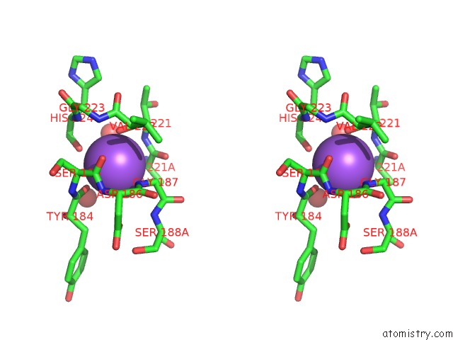
Stereo pair view
Mono view
Stereo pair view
A full contact list of Sodium with other atoms in the Na binding
site number 1 of Crystal Structure of Dfpr-Viia/Stf within 5.0Å range:
|
Reference:
S.P.Bajaj,
A.E.Schmidt,
S.Agah,
M.S.Bajaj,
K.Padmanabhan.
High Resolution Structures of P-Aminobenzamidine- and Benzamidine-Viia/Soluble Tissue Factor: Unpredicted Conformation of the 192-193 Peptide Bond and Mapping of CA2+, MG2+, Na+ and ZN2+ Sites in Factor Viia J.Biol.Chem. V. 281 24873 2006.
ISSN: ISSN 0021-9258
PubMed: 16757484
DOI: 10.1074/JBC.M509971200
Page generated: Mon Oct 7 02:27:16 2024
ISSN: ISSN 0021-9258
PubMed: 16757484
DOI: 10.1074/JBC.M509971200
Last articles
Cl in 5SHMCl in 5SHG
Cl in 5SGZ
Cl in 5SGY
Cl in 5SGR
Cl in 5SGB
Cl in 5SGG
Cl in 5SFP
Cl in 5SFG
Cl in 5SF7