Sodium »
PDB 2ekb-2fm1 »
2epf »
Sodium in PDB 2epf: Crystal Structure of Zinc-Bound Pseudecin From Pseudechis Porphyriacus
Protein crystallography data
The structure of Crystal Structure of Zinc-Bound Pseudecin From Pseudechis Porphyriacus, PDB code: 2epf
was solved by
N.Suzuki,
Y.Yamazaki,
Z.Fujimoto,
T.Morita,
H.Mizuno,
with X-Ray Crystallography technique. A brief refinement statistics is given in the table below:
| Resolution Low / High (Å) | 38.72 / 2.30 |
| Space group | P 21 21 21 |
| Cell size a, b, c (Å), α, β, γ (°) | 60.077, 62.344, 246.943, 90.00, 90.00, 90.00 |
| R / Rfree (%) | 21.9 / 27.7 |
Other elements in 2epf:
The structure of Crystal Structure of Zinc-Bound Pseudecin From Pseudechis Porphyriacus also contains other interesting chemical elements:
| Zinc | (Zn) | 5 atoms |
Sodium Binding Sites:
The binding sites of Sodium atom in the Crystal Structure of Zinc-Bound Pseudecin From Pseudechis Porphyriacus
(pdb code 2epf). This binding sites where shown within
5.0 Angstroms radius around Sodium atom.
In total 3 binding sites of Sodium where determined in the Crystal Structure of Zinc-Bound Pseudecin From Pseudechis Porphyriacus, PDB code: 2epf:
Jump to Sodium binding site number: 1; 2; 3;
In total 3 binding sites of Sodium where determined in the Crystal Structure of Zinc-Bound Pseudecin From Pseudechis Porphyriacus, PDB code: 2epf:
Jump to Sodium binding site number: 1; 2; 3;
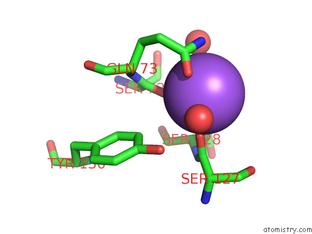
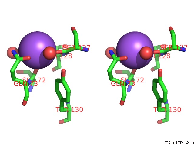
Sodium binding site 1 out of 3 in 2epf
Go back to
Sodium binding site 1 out
of 3 in the Crystal Structure of Zinc-Bound Pseudecin From Pseudechis Porphyriacus
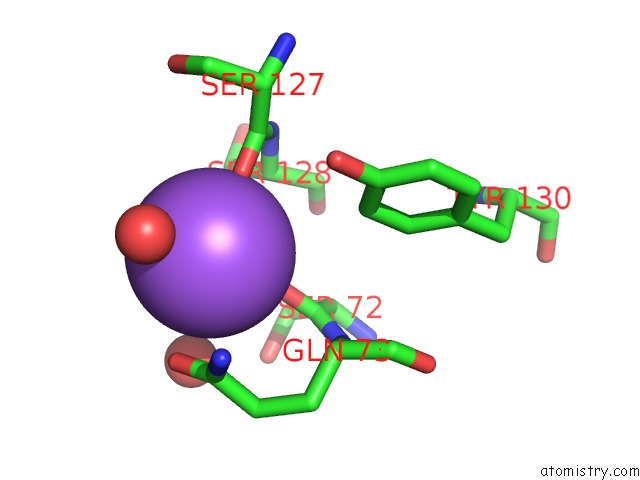
Mono view
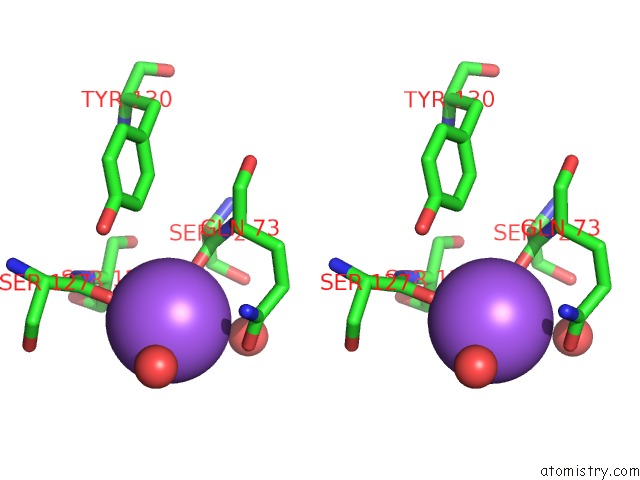
Stereo pair view
Mono view
Stereo pair view
A full contact list of Sodium with other atoms in the Na binding
site number 1 of Crystal Structure of Zinc-Bound Pseudecin From Pseudechis Porphyriacus within 5.0Å range:
|
Sodium binding site 2 out of 3 in 2epf
Go back to
Sodium binding site 2 out
of 3 in the Crystal Structure of Zinc-Bound Pseudecin From Pseudechis Porphyriacus
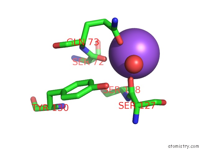
Mono view
Stereo pair view
Mono view
Stereo pair view
A full contact list of Sodium with other atoms in the Na binding
site number 2 of Crystal Structure of Zinc-Bound Pseudecin From Pseudechis Porphyriacus within 5.0Å range:
|
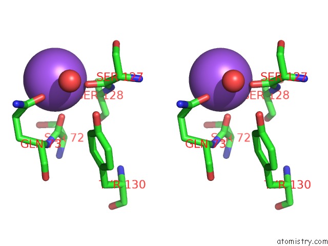
Sodium binding site 3 out of 3 in 2epf
Go back to
Sodium binding site 3 out
of 3 in the Crystal Structure of Zinc-Bound Pseudecin From Pseudechis Porphyriacus
Mono view
Stereo pair view
Mono view
Stereo pair view
A full contact list of Sodium with other atoms in the Na binding
site number 3 of Crystal Structure of Zinc-Bound Pseudecin From Pseudechis Porphyriacus within 5.0Å range:
|
Reference:
N.Suzuki,
Y.Yamazaki,
R.L.Brown,
Z.Fujimoto,
T.Morita,
H.Mizuno.
Structures of Pseudechetoxin and Pseudecin, Two Snake-Venom Cysteine-Rich Secretory Proteins That Target Cyclic Nucleotide-Gated Ion Channels: Implications For Movement of the C-Terminal Cysteine-Rich Domain Acta Crystallogr.,Sect.D V. 64 1034 2008.
ISSN: ISSN 0907-4449
PubMed: 18931410
DOI: 10.1107/S0907444908023512
Page generated: Mon Oct 7 02:24:38 2024
ISSN: ISSN 0907-4449
PubMed: 18931410
DOI: 10.1107/S0907444908023512
Last articles
Zn in 9MJ5Zn in 9HNW
Zn in 9G0L
Zn in 9FNE
Zn in 9DZN
Zn in 9E0I
Zn in 9D32
Zn in 9DAK
Zn in 8ZXC
Zn in 8ZUF