Sodium »
PDB 2e54-2eka »
2eib »
Sodium in PDB 2eib: Crystal Structure of Galactose Oxidase, W290H Mutant
Enzymatic activity of Crystal Structure of Galactose Oxidase, W290H Mutant
All present enzymatic activity of Crystal Structure of Galactose Oxidase, W290H Mutant:
1.1.3.9;
1.1.3.9;
Protein crystallography data
The structure of Crystal Structure of Galactose Oxidase, W290H Mutant, PDB code: 2eib
was solved by
S.E.Phillips,
M.J.Mcpherson,
P.F.Knowles,
C.Wilmot,
with X-Ray Crystallography technique. A brief refinement statistics is given in the table below:
| Resolution Low / High (Å) | 10.00 / 2.10 |
| Space group | P 1 21 1 |
| Cell size a, b, c (Å), α, β, γ (°) | 98.000, 89.400, 86.700, 90.00, 117.80, 90.00 |
| R / Rfree (%) | n/a / n/a |
Other elements in 2eib:
The structure of Crystal Structure of Galactose Oxidase, W290H Mutant also contains other interesting chemical elements:
| Copper | (Cu) | 1 atom |
Sodium Binding Sites:
The binding sites of Sodium atom in the Crystal Structure of Galactose Oxidase, W290H Mutant
(pdb code 2eib). This binding sites where shown within
5.0 Angstroms radius around Sodium atom.
In total only one binding site of Sodium was determined in the Crystal Structure of Galactose Oxidase, W290H Mutant, PDB code: 2eib:
In total only one binding site of Sodium was determined in the Crystal Structure of Galactose Oxidase, W290H Mutant, PDB code: 2eib:
Sodium binding site 1 out of 1 in 2eib
Go back to
Sodium binding site 1 out
of 1 in the Crystal Structure of Galactose Oxidase, W290H Mutant
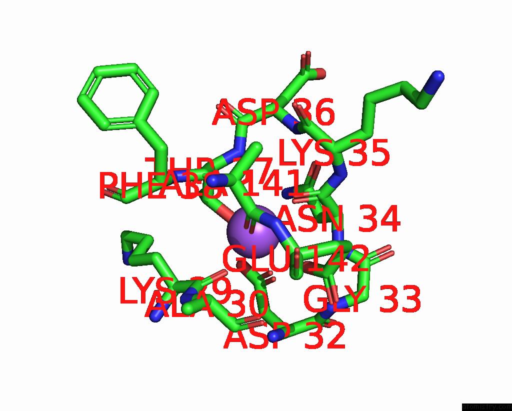
Mono view
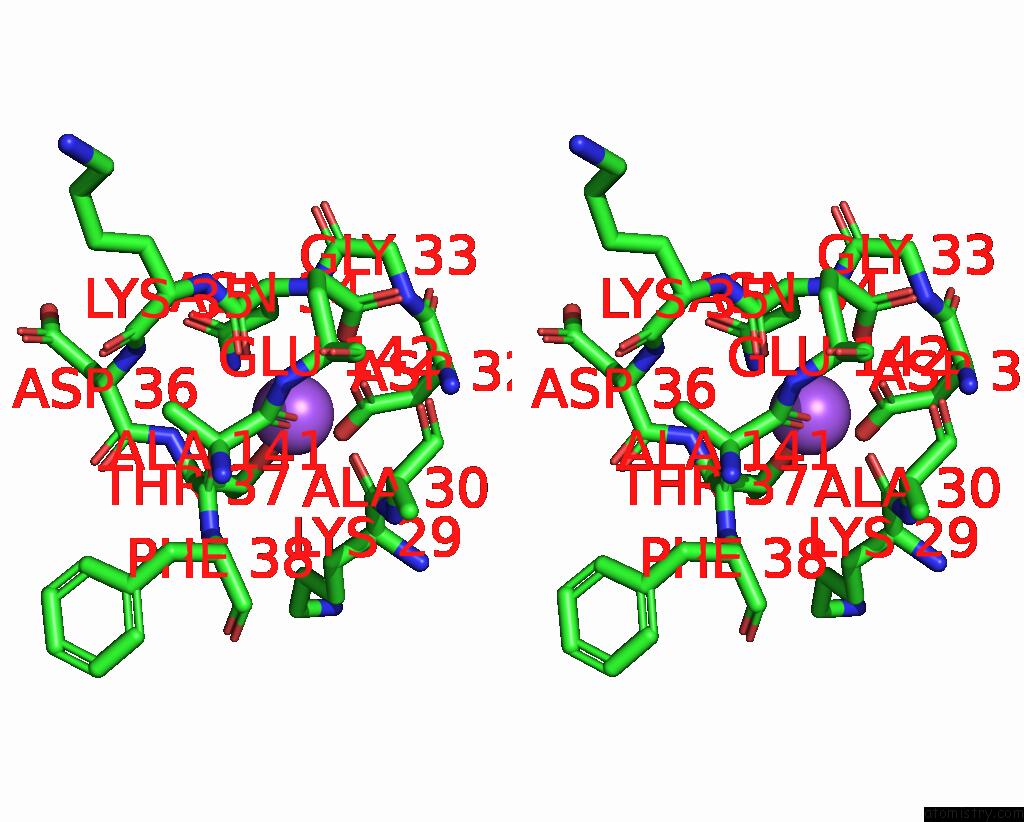
Stereo pair view
Mono view
Stereo pair view
A full contact list of Sodium with other atoms in the Na binding
site number 1 of Crystal Structure of Galactose Oxidase, W290H Mutant within 5.0Å range:
|
Reference:
M.S.Rogers,
E.M.Tyler,
N.Akyumani,
C.R.Kurtis,
R.K.Spooner,
S.E.Deacon,
S.Tamber,
S.J.Firbank,
K.Mahmoud,
P.F.Knowles,
S.E.Phillips,
M.J.Mcpherson,
D.M.Dooley.
The Stacking Tryptophan of Galactose Oxidase: A Second-Coordination Sphere Residue That Has Profound Effects on Tyrosyl Radical Behavior and Enzyme Catalysis Biochemistry V. 46 4606 2007.
ISSN: ISSN 0006-2960
PubMed: 17385891
DOI: 10.1021/BI062139D
Page generated: Mon Oct 7 02:19:05 2024
ISSN: ISSN 0006-2960
PubMed: 17385891
DOI: 10.1021/BI062139D
Last articles
Zn in 9MJ5Zn in 9HNW
Zn in 9G0L
Zn in 9FNE
Zn in 9DZN
Zn in 9E0I
Zn in 9D32
Zn in 9DAK
Zn in 8ZXC
Zn in 8ZUF