Sodium »
PDB 2czs-2e4r »
2d1e »
Sodium in PDB 2d1e: Crystal Structure of Pcya-Biliverdin Complex
Enzymatic activity of Crystal Structure of Pcya-Biliverdin Complex
All present enzymatic activity of Crystal Structure of Pcya-Biliverdin Complex:
1.3.7.5;
1.3.7.5;
Protein crystallography data
The structure of Crystal Structure of Pcya-Biliverdin Complex, PDB code: 2d1e
was solved by
Y.Hagiwara,
M.Sugishima,
Y.Takahashi,
K.Fukuyama,
with X-Ray Crystallography technique. A brief refinement statistics is given in the table below:
| Resolution Low / High (Å) | 47.67 / 1.51 |
| Space group | P 21 21 2 |
| Cell size a, b, c (Å), α, β, γ (°) | 70.826, 94.997, 42.675, 90.00, 90.00, 90.00 |
| R / Rfree (%) | 15.7 / 18.3 |
Sodium Binding Sites:
The binding sites of Sodium atom in the Crystal Structure of Pcya-Biliverdin Complex
(pdb code 2d1e). This binding sites where shown within
5.0 Angstroms radius around Sodium atom.
In total only one binding site of Sodium was determined in the Crystal Structure of Pcya-Biliverdin Complex, PDB code: 2d1e:
In total only one binding site of Sodium was determined in the Crystal Structure of Pcya-Biliverdin Complex, PDB code: 2d1e:
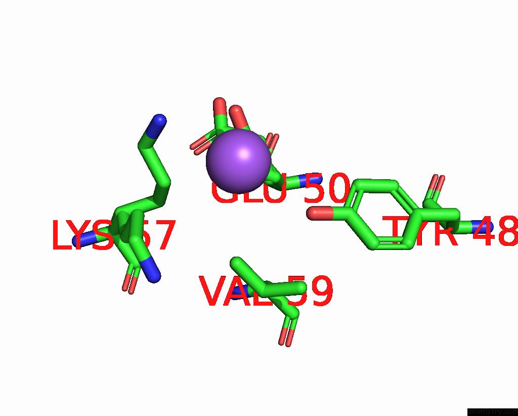
Sodium binding site 1 out of 1 in 2d1e
Go back to
Sodium binding site 1 out
of 1 in the Crystal Structure of Pcya-Biliverdin Complex
Mono view

Stereo pair view
Mono view
Stereo pair view
A full contact list of Sodium with other atoms in the Na binding
site number 1 of Crystal Structure of Pcya-Biliverdin Complex within 5.0Å range:
|
Reference:
Y.Hagiwara,
M.Sugishima,
Y.Takahashi,
K.Fukuyama.
Crystal Structure of Phycocyanobilin:Ferredoxin Oxidoreductase in Complex with Biliverdin Ixalpha, A Key Enzyme in the Biosynthesis of Phycocyanobilin Proc.Natl.Acad.Sci.Usa V. 103 27 2006.
ISSN: ISSN 0027-8424
PubMed: 16380422
DOI: 10.1073/PNAS.0507266103
Page generated: Mon Oct 7 02:11:03 2024
ISSN: ISSN 0027-8424
PubMed: 16380422
DOI: 10.1073/PNAS.0507266103
Last articles
Cl in 5EGMCl in 5EFF
Cl in 5EEI
Cl in 5EEN
Cl in 5EFD
Cl in 5EE5
Cl in 5EEK
Cl in 5EE3
Cl in 5EE7
Cl in 5EDI