Sodium »
PDB 2aoe-2bhp »
2bfg »
Sodium in PDB 2bfg: Crystal Structure of Beta-Xylosidase (Fam GH39) in Complex with Dinitrophenyl-Beta-Xyloside and Covalently Bound Xyloside
Enzymatic activity of Crystal Structure of Beta-Xylosidase (Fam GH39) in Complex with Dinitrophenyl-Beta-Xyloside and Covalently Bound Xyloside
All present enzymatic activity of Crystal Structure of Beta-Xylosidase (Fam GH39) in Complex with Dinitrophenyl-Beta-Xyloside and Covalently Bound Xyloside:
3.2.1.37;
3.2.1.37;
Protein crystallography data
The structure of Crystal Structure of Beta-Xylosidase (Fam GH39) in Complex with Dinitrophenyl-Beta-Xyloside and Covalently Bound Xyloside, PDB code: 2bfg
was solved by
M.Czjzek,
T.Bravman,
B.Henrissat,
Y.Shoham,
with X-Ray Crystallography technique. A brief refinement statistics is given in the table below:
| Resolution Low / High (Å) | 40.11 / 2.40 |
| Space group | P 21 21 21 |
| Cell size a, b, c (Å), α, β, γ (°) | 88.950, 162.160, 308.200, 90.00, 90.00, 90.00 |
| R / Rfree (%) | 19.4 / 27 |
Sodium Binding Sites:
The binding sites of Sodium atom in the Crystal Structure of Beta-Xylosidase (Fam GH39) in Complex with Dinitrophenyl-Beta-Xyloside and Covalently Bound Xyloside
(pdb code 2bfg). This binding sites where shown within
5.0 Angstroms radius around Sodium atom.
In total 8 binding sites of Sodium where determined in the Crystal Structure of Beta-Xylosidase (Fam GH39) in Complex with Dinitrophenyl-Beta-Xyloside and Covalently Bound Xyloside, PDB code: 2bfg:
Jump to Sodium binding site number: 1; 2; 3; 4; 5; 6; 7; 8;
In total 8 binding sites of Sodium where determined in the Crystal Structure of Beta-Xylosidase (Fam GH39) in Complex with Dinitrophenyl-Beta-Xyloside and Covalently Bound Xyloside, PDB code: 2bfg:
Jump to Sodium binding site number: 1; 2; 3; 4; 5; 6; 7; 8;
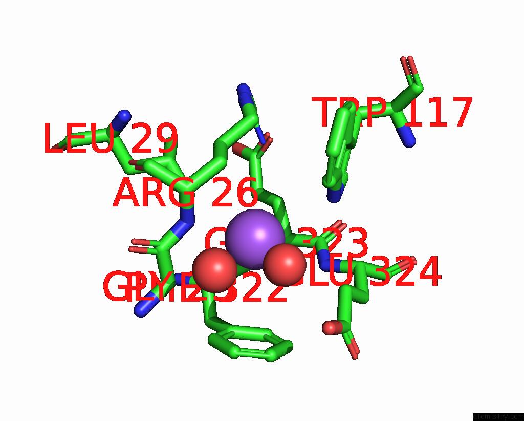
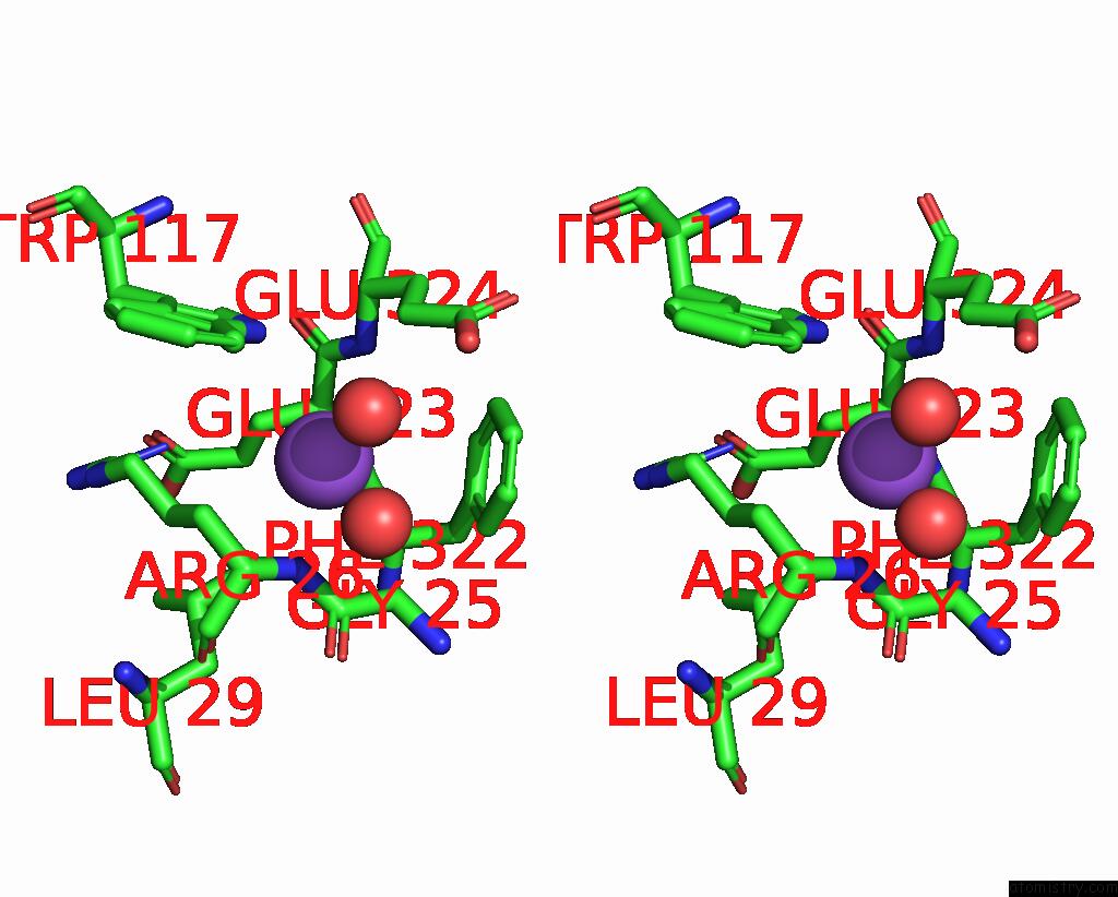
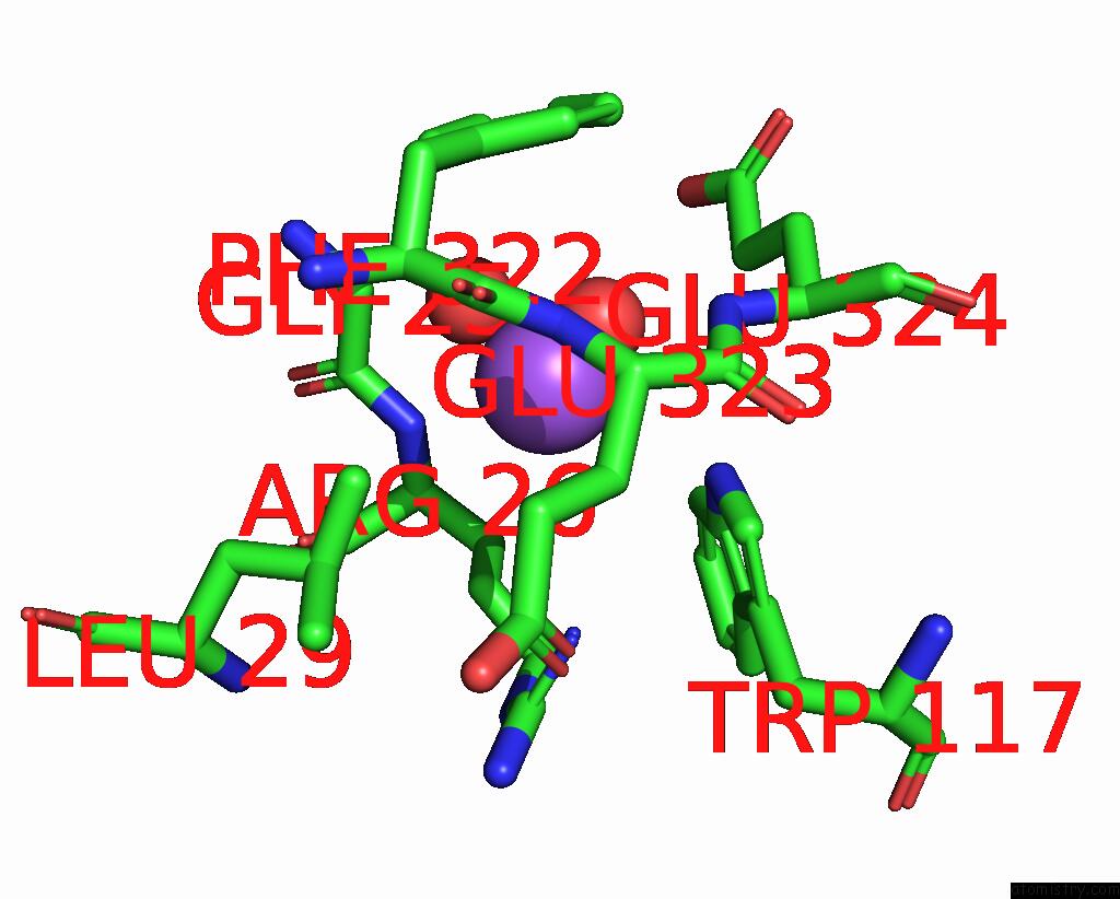
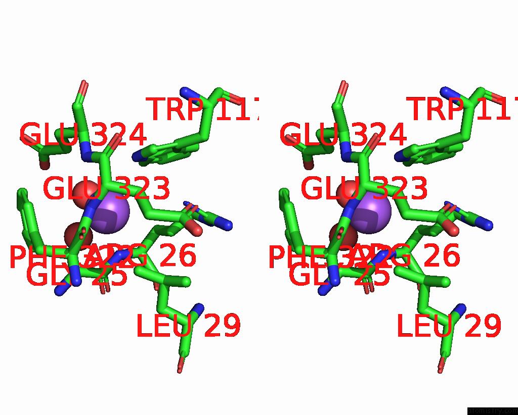
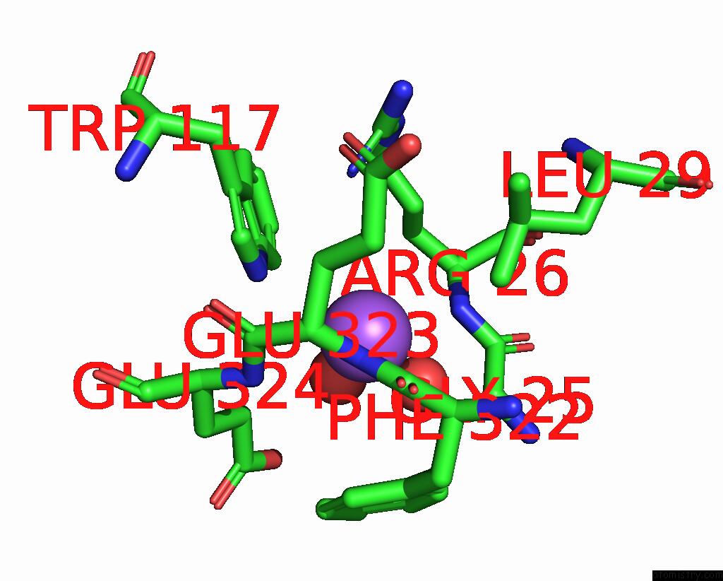
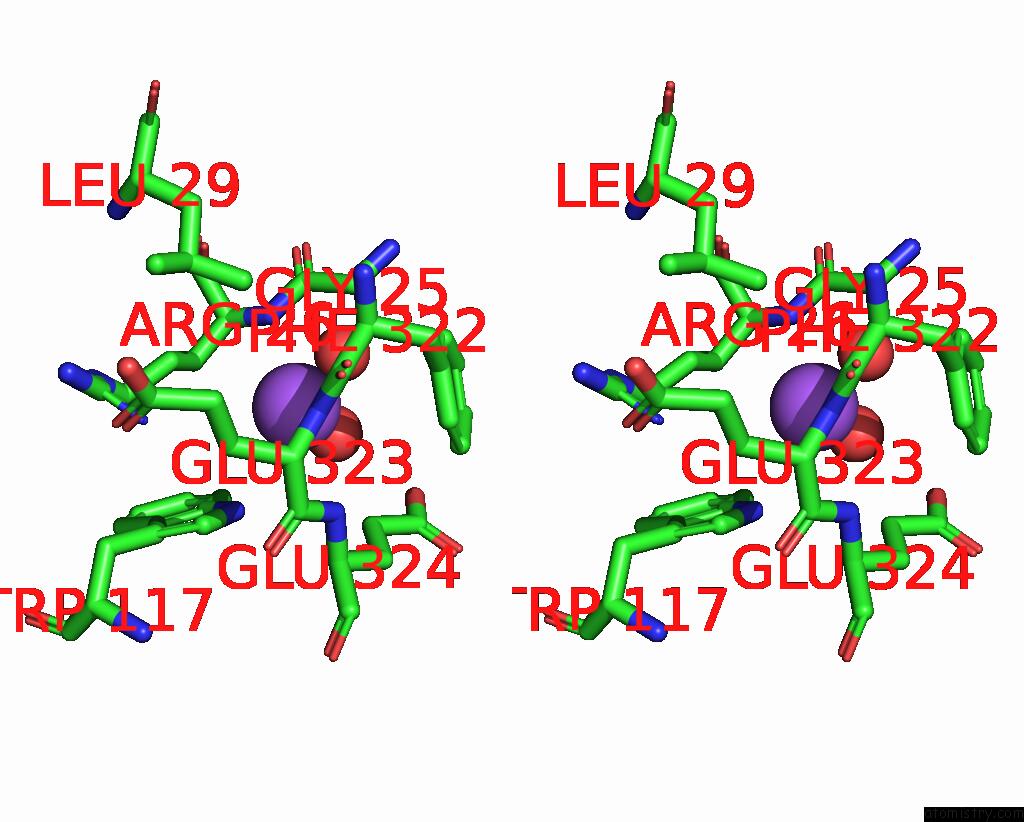
Sodium binding site 1 out of 8 in 2bfg
Go back to
Sodium binding site 1 out
of 8 in the Crystal Structure of Beta-Xylosidase (Fam GH39) in Complex with Dinitrophenyl-Beta-Xyloside and Covalently Bound Xyloside
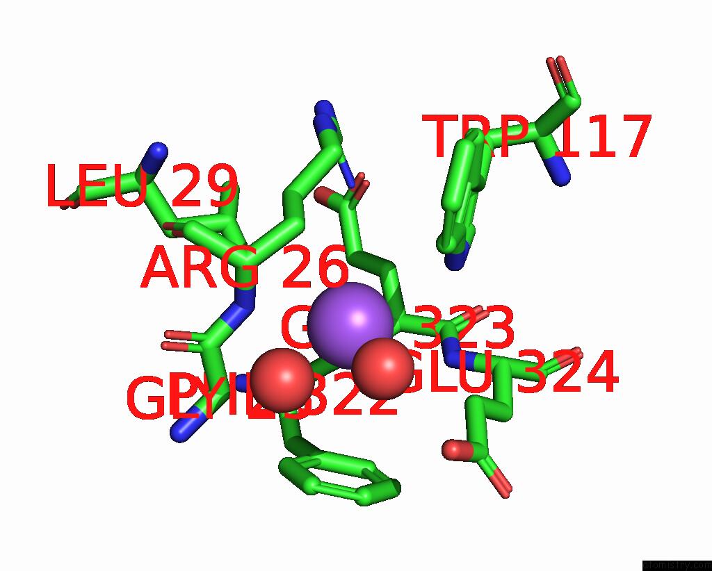
Mono view
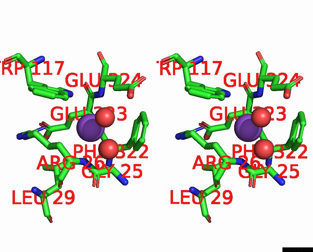
Stereo pair view
Mono view
Stereo pair view
A full contact list of Sodium with other atoms in the Na binding
site number 1 of Crystal Structure of Beta-Xylosidase (Fam GH39) in Complex with Dinitrophenyl-Beta-Xyloside and Covalently Bound Xyloside within 5.0Å range:
|
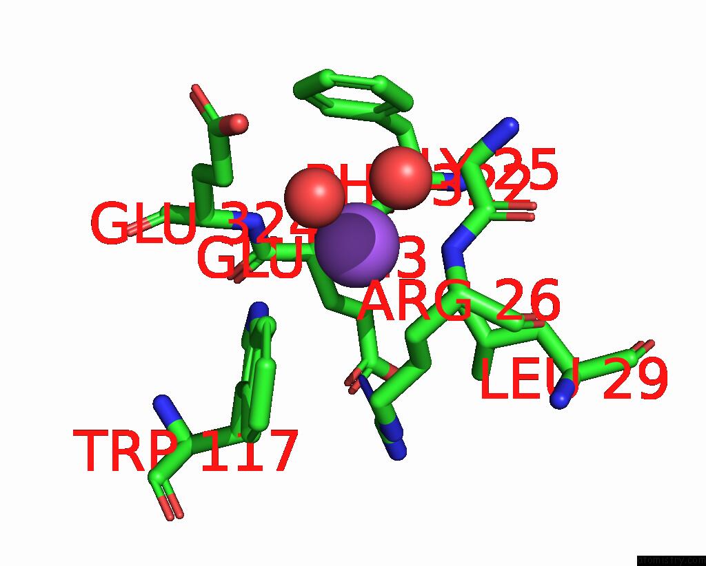
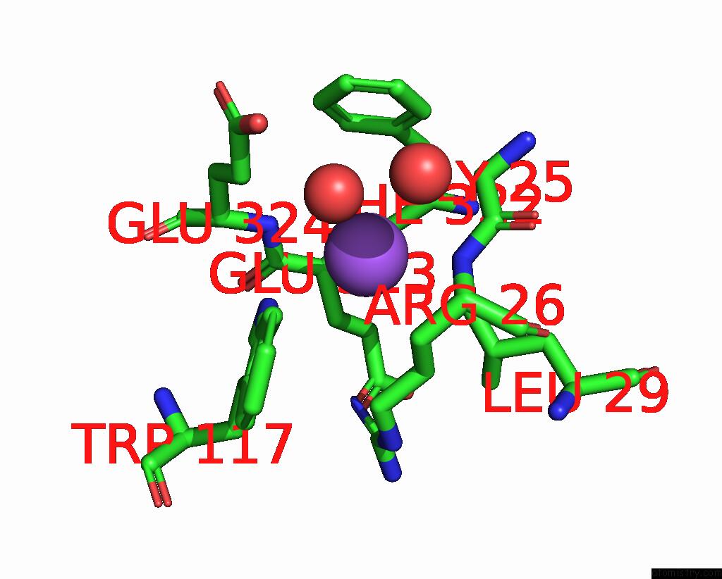
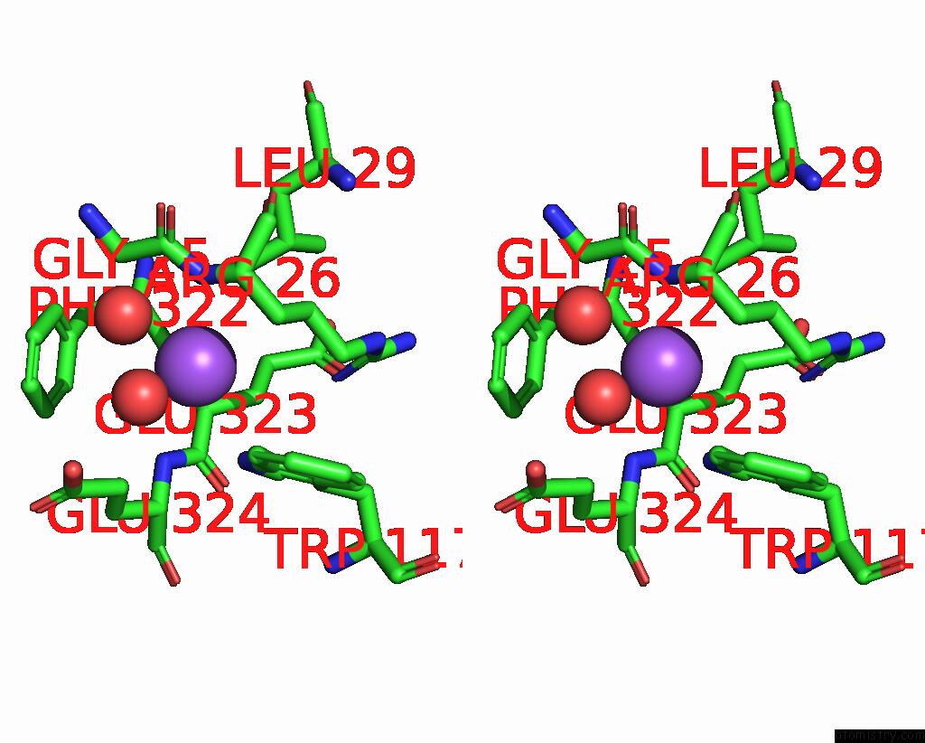
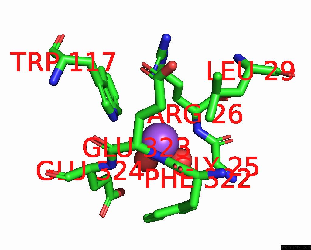
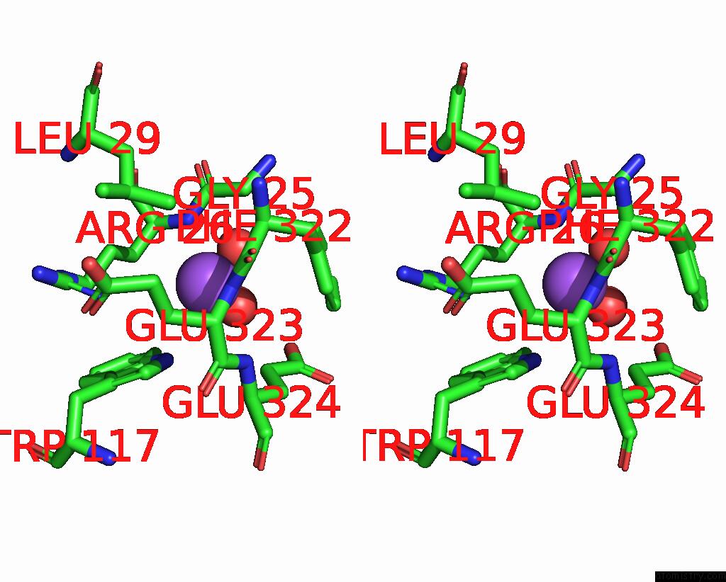
Sodium binding site 2 out of 8 in 2bfg
Go back to
Sodium binding site 2 out
of 8 in the Crystal Structure of Beta-Xylosidase (Fam GH39) in Complex with Dinitrophenyl-Beta-Xyloside and Covalently Bound Xyloside
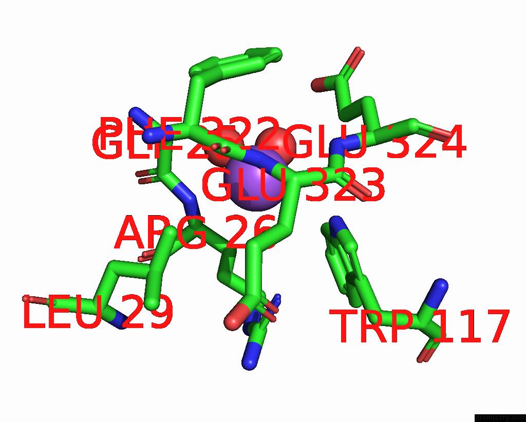
Mono view
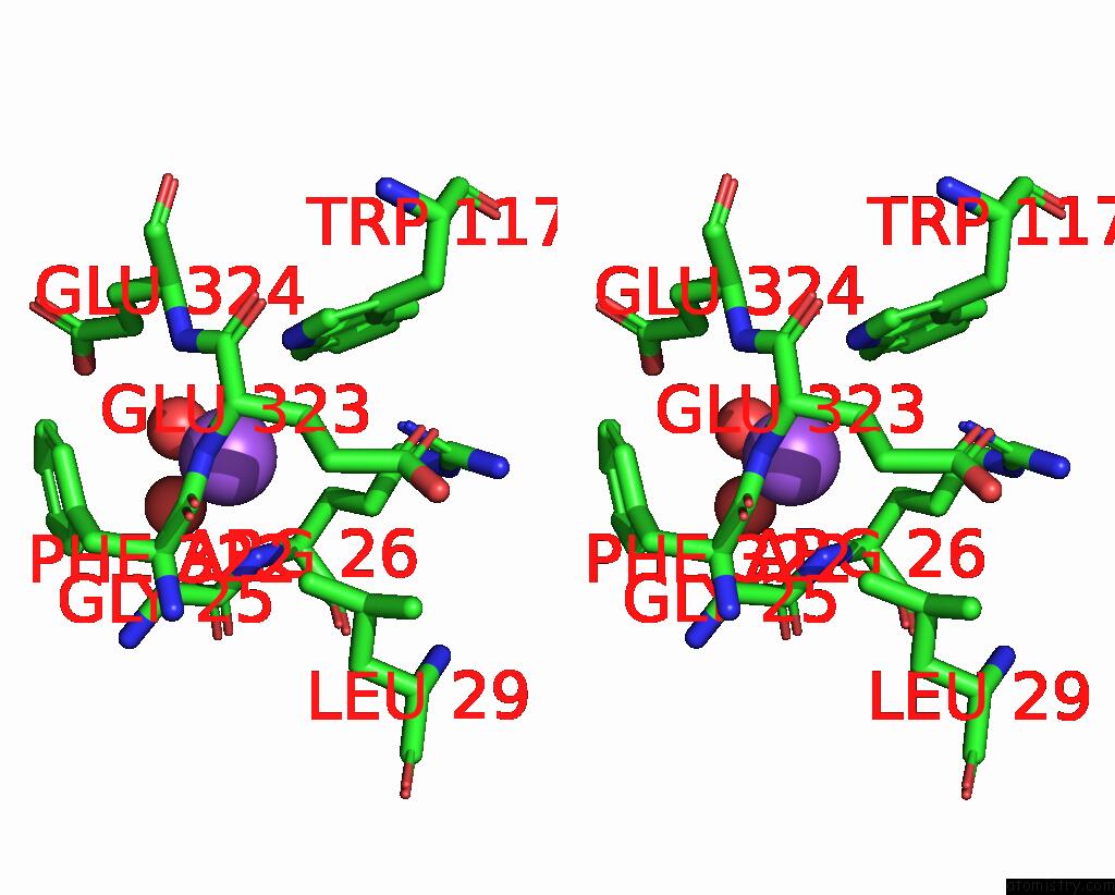
Stereo pair view
Mono view
Stereo pair view
A full contact list of Sodium with other atoms in the Na binding
site number 2 of Crystal Structure of Beta-Xylosidase (Fam GH39) in Complex with Dinitrophenyl-Beta-Xyloside and Covalently Bound Xyloside within 5.0Å range:
|
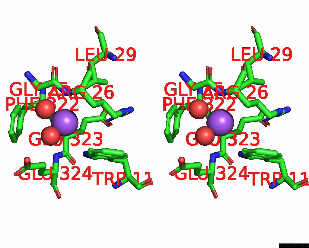
Sodium binding site 3 out of 8 in 2bfg
Go back to
Sodium binding site 3 out
of 8 in the Crystal Structure of Beta-Xylosidase (Fam GH39) in Complex with Dinitrophenyl-Beta-Xyloside and Covalently Bound Xyloside
Mono view
Stereo pair view
Mono view
Stereo pair view
A full contact list of Sodium with other atoms in the Na binding
site number 3 of Crystal Structure of Beta-Xylosidase (Fam GH39) in Complex with Dinitrophenyl-Beta-Xyloside and Covalently Bound Xyloside within 5.0Å range:
|
Sodium binding site 4 out of 8 in 2bfg
Go back to
Sodium binding site 4 out
of 8 in the Crystal Structure of Beta-Xylosidase (Fam GH39) in Complex with Dinitrophenyl-Beta-Xyloside and Covalently Bound Xyloside
Mono view
Stereo pair view
Mono view
Stereo pair view
A full contact list of Sodium with other atoms in the Na binding
site number 4 of Crystal Structure of Beta-Xylosidase (Fam GH39) in Complex with Dinitrophenyl-Beta-Xyloside and Covalently Bound Xyloside within 5.0Å range:
|
Sodium binding site 5 out of 8 in 2bfg
Go back to
Sodium binding site 5 out
of 8 in the Crystal Structure of Beta-Xylosidase (Fam GH39) in Complex with Dinitrophenyl-Beta-Xyloside and Covalently Bound Xyloside
Mono view
Stereo pair view
Mono view
Stereo pair view
A full contact list of Sodium with other atoms in the Na binding
site number 5 of Crystal Structure of Beta-Xylosidase (Fam GH39) in Complex with Dinitrophenyl-Beta-Xyloside and Covalently Bound Xyloside within 5.0Å range:
|
Sodium binding site 6 out of 8 in 2bfg
Go back to
Sodium binding site 6 out
of 8 in the Crystal Structure of Beta-Xylosidase (Fam GH39) in Complex with Dinitrophenyl-Beta-Xyloside and Covalently Bound Xyloside
Mono view
Stereo pair view
Mono view
Stereo pair view
A full contact list of Sodium with other atoms in the Na binding
site number 6 of Crystal Structure of Beta-Xylosidase (Fam GH39) in Complex with Dinitrophenyl-Beta-Xyloside and Covalently Bound Xyloside within 5.0Å range:
|
Sodium binding site 7 out of 8 in 2bfg
Go back to
Sodium binding site 7 out
of 8 in the Crystal Structure of Beta-Xylosidase (Fam GH39) in Complex with Dinitrophenyl-Beta-Xyloside and Covalently Bound Xyloside
Mono view
Stereo pair view
Mono view
Stereo pair view
A full contact list of Sodium with other atoms in the Na binding
site number 7 of Crystal Structure of Beta-Xylosidase (Fam GH39) in Complex with Dinitrophenyl-Beta-Xyloside and Covalently Bound Xyloside within 5.0Å range:
|
Sodium binding site 8 out of 8 in 2bfg
Go back to
Sodium binding site 8 out
of 8 in the Crystal Structure of Beta-Xylosidase (Fam GH39) in Complex with Dinitrophenyl-Beta-Xyloside and Covalently Bound Xyloside
Mono view
Stereo pair view
Mono view
Stereo pair view
A full contact list of Sodium with other atoms in the Na binding
site number 8 of Crystal Structure of Beta-Xylosidase (Fam GH39) in Complex with Dinitrophenyl-Beta-Xyloside and Covalently Bound Xyloside within 5.0Å range:
|
Reference:
M.Czjzek,
A.B.David,
T.Bravman,
G.Shoham,
B.Henrissat,
Y.Shoham.
Enzyme-Substrate Complex Structures of A GH39 Beta- Xylosidase From Geobacillus Stearothermophilus. J.Mol.Biol. V. 353 838 2005.
ISSN: ISSN 0022-2836
PubMed: 16212978
DOI: 10.1016/J.JMB.2005.09.003
Page generated: Sun Aug 17 10:10:36 2025
ISSN: ISSN 0022-2836
PubMed: 16212978
DOI: 10.1016/J.JMB.2005.09.003
Last articles
Na in 4OFXNa in 4OE2
Na in 4OEH
Na in 4ODI
Na in 4OF4
Na in 4ODN
Na in 4OCI
Na in 4OCJ
Na in 4OBO
Na in 4O7J