Sodium »
PDB 1zum-2aoc »
2ahs »
Sodium in PDB 2ahs: Crystal Structure of the Catalytic Domain of Human Tyrosine Receptor Phosphatase Beta
Enzymatic activity of Crystal Structure of the Catalytic Domain of Human Tyrosine Receptor Phosphatase Beta
All present enzymatic activity of Crystal Structure of the Catalytic Domain of Human Tyrosine Receptor Phosphatase Beta:
3.1.3.48;
3.1.3.48;
Protein crystallography data
The structure of Crystal Structure of the Catalytic Domain of Human Tyrosine Receptor Phosphatase Beta, PDB code: 2ahs
was solved by
E.Ugochukwu,
J.Eswaran,
A.Barr,
O.Gileadi,
F.Sobott,
N.Burgess,
L.Ball,
J.Bray,
F.Von Delft,
J.Debreczeni,
G.Bunkoczi,
A.Turnbull,
S.Das,
J.Weigelt,
A.Edwards,
C.Arrowsmith,
M.Sundstrom,
S.Knapp,
Structuralgenomics Consortium (Sgc),
with X-Ray Crystallography technique. A brief refinement statistics is given in the table below:
| Resolution Low / High (Å) | 53.40 / 2.10 |
| Space group | P 61 2 2 |
| Cell size a, b, c (Å), α, β, γ (°) | 123.368, 123.368, 179.569, 90.00, 90.00, 120.00 |
| R / Rfree (%) | 15.2 / 20.6 |
Other elements in 2ahs:
The structure of Crystal Structure of the Catalytic Domain of Human Tyrosine Receptor Phosphatase Beta also contains other interesting chemical elements:
| Chlorine | (Cl) | 9 atoms |
Sodium Binding Sites:
The binding sites of Sodium atom in the Crystal Structure of the Catalytic Domain of Human Tyrosine Receptor Phosphatase Beta
(pdb code 2ahs). This binding sites where shown within
5.0 Angstroms radius around Sodium atom.
In total only one binding site of Sodium was determined in the Crystal Structure of the Catalytic Domain of Human Tyrosine Receptor Phosphatase Beta, PDB code: 2ahs:
In total only one binding site of Sodium was determined in the Crystal Structure of the Catalytic Domain of Human Tyrosine Receptor Phosphatase Beta, PDB code: 2ahs:
Sodium binding site 1 out of 1 in 2ahs
Go back to
Sodium binding site 1 out
of 1 in the Crystal Structure of the Catalytic Domain of Human Tyrosine Receptor Phosphatase Beta
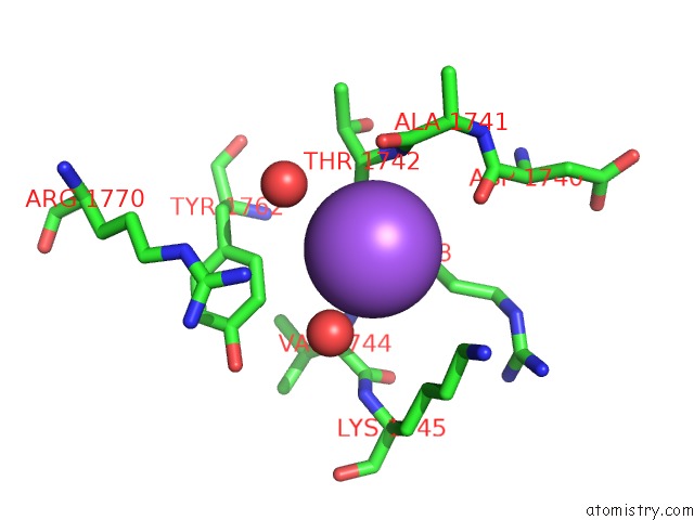
Mono view
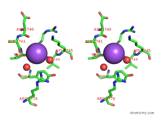
Stereo pair view
Mono view
Stereo pair view
A full contact list of Sodium with other atoms in the Na binding
site number 1 of Crystal Structure of the Catalytic Domain of Human Tyrosine Receptor Phosphatase Beta within 5.0Å range:
|
Reference:
A.J.Barr,
E.Ugochukwu,
W.H.Lee,
O.N.King,
P.Filippakopoulos,
I.Alfano,
P.Savitsky,
N.A.Burgess-Brown,
S.Muller,
S.Knapp.
Large-Scale Structural Analysis of the Classical Human Protein Tyrosine Phosphatome. Cell(Cambridge,Mass.) V. 136 352 2009.
ISSN: ISSN 0092-8674
PubMed: 19167335
DOI: 10.1016/J.CELL.2008.11.038
Page generated: Mon Oct 7 01:52:14 2024
ISSN: ISSN 0092-8674
PubMed: 19167335
DOI: 10.1016/J.CELL.2008.11.038
Last articles
Fe in 2YXOFe in 2YRS
Fe in 2YXC
Fe in 2YNM
Fe in 2YVJ
Fe in 2YP1
Fe in 2YU2
Fe in 2YU1
Fe in 2YQB
Fe in 2YOO