Sodium »
PDB 1zum-2aoc »
2abs »
Sodium in PDB 2abs: Crystal Structure of T. Gondii Adenosine Kinase Complexed with Amp-Pcp
Enzymatic activity of Crystal Structure of T. Gondii Adenosine Kinase Complexed with Amp-Pcp
All present enzymatic activity of Crystal Structure of T. Gondii Adenosine Kinase Complexed with Amp-Pcp:
2.7.1.20;
2.7.1.20;
Protein crystallography data
The structure of Crystal Structure of T. Gondii Adenosine Kinase Complexed with Amp-Pcp, PDB code: 2abs
was solved by
Y.Zhang,
M.H.El Kouni,
S.E.Ealick,
with X-Ray Crystallography technique. A brief refinement statistics is given in the table below:
| Resolution Low / High (Å) | 10.00 / 1.10 |
| Space group | P 21 21 21 |
| Cell size a, b, c (Å), α, β, γ (°) | 60.051, 67.923, 83.192, 90.00, 90.00, 90.00 |
| R / Rfree (%) | 13.5 / 16.7 |
Other elements in 2abs:
The structure of Crystal Structure of T. Gondii Adenosine Kinase Complexed with Amp-Pcp also contains other interesting chemical elements:
| Chlorine | (Cl) | 1 atom |
Sodium Binding Sites:
The binding sites of Sodium atom in the Crystal Structure of T. Gondii Adenosine Kinase Complexed with Amp-Pcp
(pdb code 2abs). This binding sites where shown within
5.0 Angstroms radius around Sodium atom.
In total only one binding site of Sodium was determined in the Crystal Structure of T. Gondii Adenosine Kinase Complexed with Amp-Pcp, PDB code: 2abs:
In total only one binding site of Sodium was determined in the Crystal Structure of T. Gondii Adenosine Kinase Complexed with Amp-Pcp, PDB code: 2abs:
Sodium binding site 1 out of 1 in 2abs
Go back to
Sodium binding site 1 out
of 1 in the Crystal Structure of T. Gondii Adenosine Kinase Complexed with Amp-Pcp
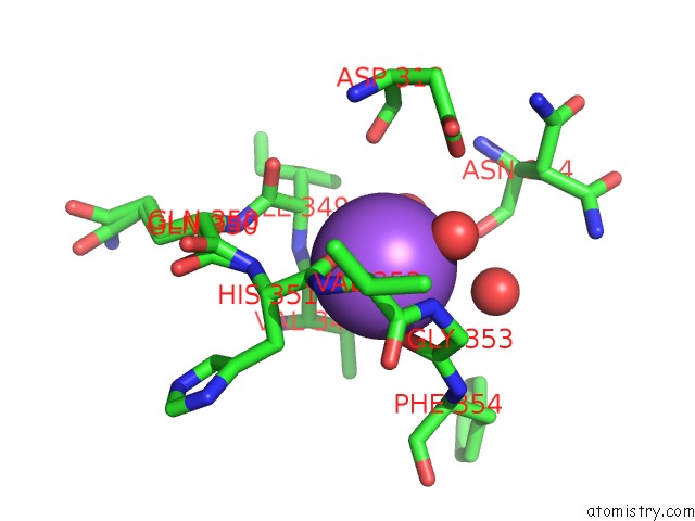
Mono view
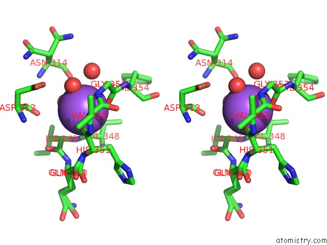
Stereo pair view
Mono view
Stereo pair view
A full contact list of Sodium with other atoms in the Na binding
site number 1 of Crystal Structure of T. Gondii Adenosine Kinase Complexed with Amp-Pcp within 5.0Å range:
|
Reference:
Y.Zhang,
M.H.El Kouni,
S.E.Ealick.
Structure of Toxoplasma Gondii Adenosine Kinase in Complex with An Atp Analog at 1.1 Angstroms Resolution. Acta Crystallogr.,Sect.D V. 62 140 2006.
ISSN: ISSN 0907-4449
PubMed: 16421444
DOI: 10.1107/S090744490503430X
Page generated: Mon Oct 7 01:51:25 2024
ISSN: ISSN 0907-4449
PubMed: 16421444
DOI: 10.1107/S090744490503430X
Last articles
Zn in 9J0NZn in 9J0O
Zn in 9J0P
Zn in 9FJX
Zn in 9EKB
Zn in 9C0F
Zn in 9CAH
Zn in 9CH0
Zn in 9CH3
Zn in 9CH1