Sodium »
PDB 1zum-2aoc »
2a9y »
Sodium in PDB 2a9y: Crystal Structure of T. Gondii Adenosine Kinase Complexed with N6- Dimethyladenosine
Enzymatic activity of Crystal Structure of T. Gondii Adenosine Kinase Complexed with N6- Dimethyladenosine
All present enzymatic activity of Crystal Structure of T. Gondii Adenosine Kinase Complexed with N6- Dimethyladenosine:
2.7.1.20;
2.7.1.20;
Protein crystallography data
The structure of Crystal Structure of T. Gondii Adenosine Kinase Complexed with N6- Dimethyladenosine, PDB code: 2a9y
was solved by
Y.Zhang,
M.H.El Kouni,
S.E.Ealick,
with X-Ray Crystallography technique. A brief refinement statistics is given in the table below:
| Resolution Low / High (Å) | 10.00 / 1.35 |
| Space group | P 21 21 21 |
| Cell size a, b, c (Å), α, β, γ (°) | 60.285, 61.516, 91.828, 90.00, 90.00, 90.00 |
| R / Rfree (%) | 13.4 / 19.6 |
Other elements in 2a9y:
The structure of Crystal Structure of T. Gondii Adenosine Kinase Complexed with N6- Dimethyladenosine also contains other interesting chemical elements:
| Chlorine | (Cl) | 1 atom |
Sodium Binding Sites:
The binding sites of Sodium atom in the Crystal Structure of T. Gondii Adenosine Kinase Complexed with N6- Dimethyladenosine
(pdb code 2a9y). This binding sites where shown within
5.0 Angstroms radius around Sodium atom.
In total 2 binding sites of Sodium where determined in the Crystal Structure of T. Gondii Adenosine Kinase Complexed with N6- Dimethyladenosine, PDB code: 2a9y:
Jump to Sodium binding site number: 1; 2;
In total 2 binding sites of Sodium where determined in the Crystal Structure of T. Gondii Adenosine Kinase Complexed with N6- Dimethyladenosine, PDB code: 2a9y:
Jump to Sodium binding site number: 1; 2;
Sodium binding site 1 out of 2 in 2a9y
Go back to
Sodium binding site 1 out
of 2 in the Crystal Structure of T. Gondii Adenosine Kinase Complexed with N6- Dimethyladenosine
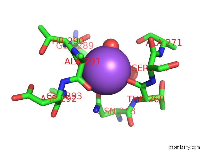
Mono view
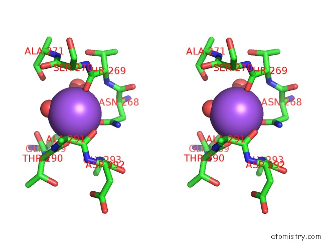
Stereo pair view
Mono view
Stereo pair view
A full contact list of Sodium with other atoms in the Na binding
site number 1 of Crystal Structure of T. Gondii Adenosine Kinase Complexed with N6- Dimethyladenosine within 5.0Å range:
|
Sodium binding site 2 out of 2 in 2a9y
Go back to
Sodium binding site 2 out
of 2 in the Crystal Structure of T. Gondii Adenosine Kinase Complexed with N6- Dimethyladenosine
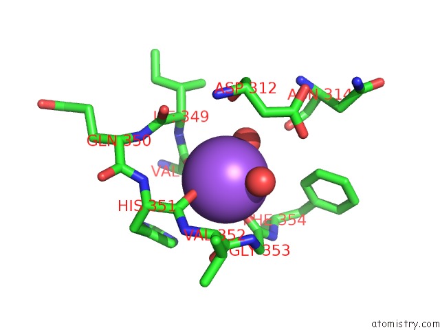
Mono view
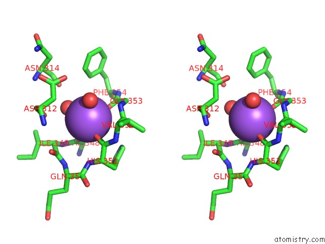
Stereo pair view
Mono view
Stereo pair view
A full contact list of Sodium with other atoms in the Na binding
site number 2 of Crystal Structure of T. Gondii Adenosine Kinase Complexed with N6- Dimethyladenosine within 5.0Å range:
|
Reference:
Y.Zhang,
M.H.El Kouni,
S.E.Ealick.
Substrate Analogs Induce An Intermediate Conformational Change in Toxoplasma Gondii Adenosine Kinase Acta Crystallogr.,Sect.D V. 63 126 2007.
ISSN: ISSN 0907-4449
PubMed: 17242506
DOI: 10.1107/S0907444906043654
Page generated: Mon Oct 7 01:49:58 2024
ISSN: ISSN 0907-4449
PubMed: 17242506
DOI: 10.1107/S0907444906043654
Last articles
Zn in 9J0NZn in 9J0O
Zn in 9J0P
Zn in 9FJX
Zn in 9EKB
Zn in 9C0F
Zn in 9CAH
Zn in 9CH0
Zn in 9CH3
Zn in 9CH1