Sodium »
PDB 1zum-2aoc »
2a2c »
Sodium in PDB 2a2c: X-Ray Structure of Human N-Acetyl Galactosamine Kinase Complexed with Mg-Adp and N-Acetyl Galactosamine 1-Phosphate
Protein crystallography data
The structure of X-Ray Structure of Human N-Acetyl Galactosamine Kinase Complexed with Mg-Adp and N-Acetyl Galactosamine 1-Phosphate, PDB code: 2a2c
was solved by
J.B.Thoden,
H.M.Holden,
with X-Ray Crystallography technique. A brief refinement statistics is given in the table below:
| Resolution Low / High (Å) | 50.00 / 1.65 |
| Space group | P 65 |
| Cell size a, b, c (Å), α, β, γ (°) | 123.800, 123.800, 60.100, 90.00, 90.00, 120.00 |
| R / Rfree (%) | n/a / n/a |
Other elements in 2a2c:
The structure of X-Ray Structure of Human N-Acetyl Galactosamine Kinase Complexed with Mg-Adp and N-Acetyl Galactosamine 1-Phosphate also contains other interesting chemical elements:
| Magnesium | (Mg) | 1 atom |
| Chlorine | (Cl) | 1 atom |
Sodium Binding Sites:
The binding sites of Sodium atom in the X-Ray Structure of Human N-Acetyl Galactosamine Kinase Complexed with Mg-Adp and N-Acetyl Galactosamine 1-Phosphate
(pdb code 2a2c). This binding sites where shown within
5.0 Angstroms radius around Sodium atom.
In total only one binding site of Sodium was determined in the X-Ray Structure of Human N-Acetyl Galactosamine Kinase Complexed with Mg-Adp and N-Acetyl Galactosamine 1-Phosphate, PDB code: 2a2c:
In total only one binding site of Sodium was determined in the X-Ray Structure of Human N-Acetyl Galactosamine Kinase Complexed with Mg-Adp and N-Acetyl Galactosamine 1-Phosphate, PDB code: 2a2c:
Sodium binding site 1 out of 1 in 2a2c
Go back to
Sodium binding site 1 out
of 1 in the X-Ray Structure of Human N-Acetyl Galactosamine Kinase Complexed with Mg-Adp and N-Acetyl Galactosamine 1-Phosphate
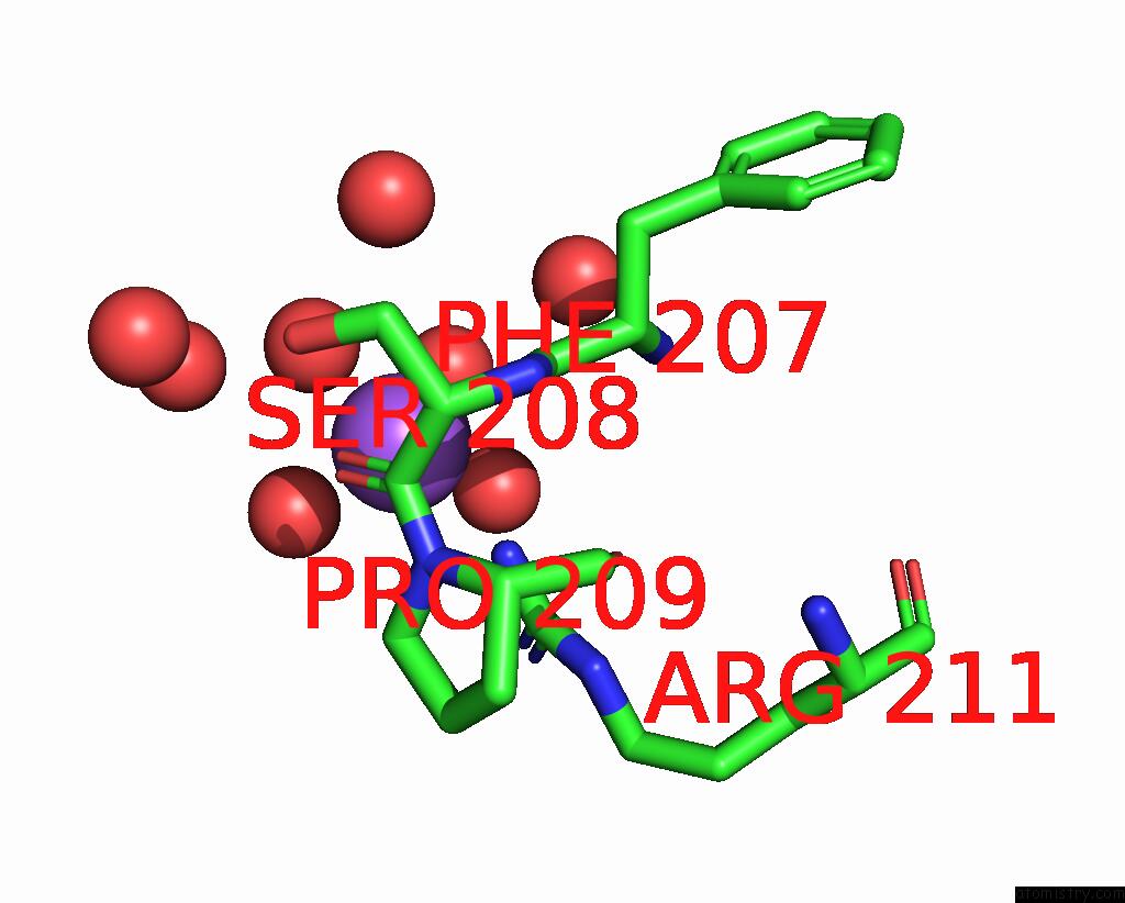
Mono view
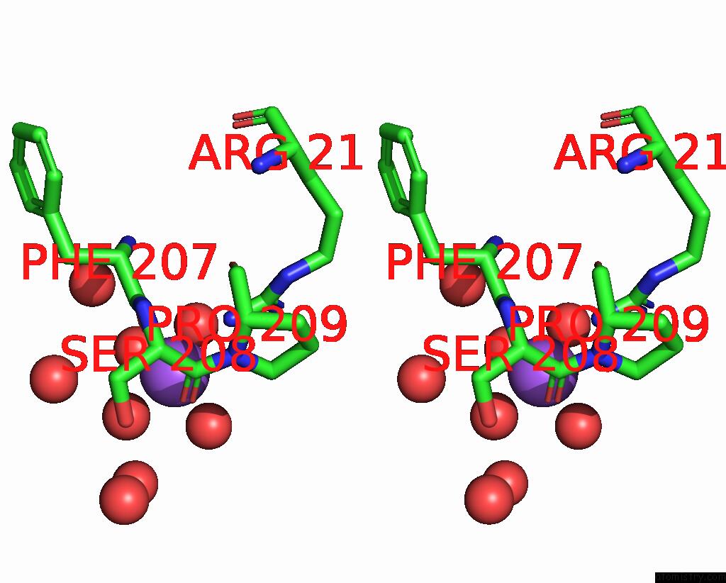
Stereo pair view
Mono view
Stereo pair view
A full contact list of Sodium with other atoms in the Na binding
site number 1 of X-Ray Structure of Human N-Acetyl Galactosamine Kinase Complexed with Mg-Adp and N-Acetyl Galactosamine 1-Phosphate within 5.0Å range:
|
Reference:
J.B.Thoden,
H.M.Holden.
The Molecular Architecture of Human N-Acetylgalactosamine Kinase. J.Biol.Chem. V. 280 32784 2005.
ISSN: ISSN 0021-9258
PubMed: 16006554
DOI: 10.1074/JBC.M505730200
Page generated: Mon Oct 7 01:47:11 2024
ISSN: ISSN 0021-9258
PubMed: 16006554
DOI: 10.1074/JBC.M505730200
Last articles
Zn in 9J0NZn in 9J0O
Zn in 9J0P
Zn in 9FJX
Zn in 9EKB
Zn in 9C0F
Zn in 9CAH
Zn in 9CH0
Zn in 9CH3
Zn in 9CH1