Sodium »
PDB 1z2u-1zud »
1zob »
Sodium in PDB 1zob: Crystal Structure of Dialkylglycine Decarboxylases Bound with Calcium Ion
Enzymatic activity of Crystal Structure of Dialkylglycine Decarboxylases Bound with Calcium Ion
All present enzymatic activity of Crystal Structure of Dialkylglycine Decarboxylases Bound with Calcium Ion:
4.1.1.64;
4.1.1.64;
Protein crystallography data
The structure of Crystal Structure of Dialkylglycine Decarboxylases Bound with Calcium Ion, PDB code: 1zob
was solved by
W.Liu,
M.D.Toney,
with X-Ray Crystallography technique. A brief refinement statistics is given in the table below:
| Resolution Low / High (Å) | 30.00 / 2.75 |
| Space group | P 64 2 2 |
| Cell size a, b, c (Å), α, β, γ (°) | 150.553, 150.553, 84.703, 90.00, 90.00, 120.00 |
| R / Rfree (%) | 21.9 / 27.7 |
Other elements in 1zob:
The structure of Crystal Structure of Dialkylglycine Decarboxylases Bound with Calcium Ion also contains other interesting chemical elements:
| Calcium | (Ca) | 1 atom |
Sodium Binding Sites:
The binding sites of Sodium atom in the Crystal Structure of Dialkylglycine Decarboxylases Bound with Calcium Ion
(pdb code 1zob). This binding sites where shown within
5.0 Angstroms radius around Sodium atom.
In total only one binding site of Sodium was determined in the Crystal Structure of Dialkylglycine Decarboxylases Bound with Calcium Ion, PDB code: 1zob:
In total only one binding site of Sodium was determined in the Crystal Structure of Dialkylglycine Decarboxylases Bound with Calcium Ion, PDB code: 1zob:
Sodium binding site 1 out of 1 in 1zob
Go back to
Sodium binding site 1 out
of 1 in the Crystal Structure of Dialkylglycine Decarboxylases Bound with Calcium Ion
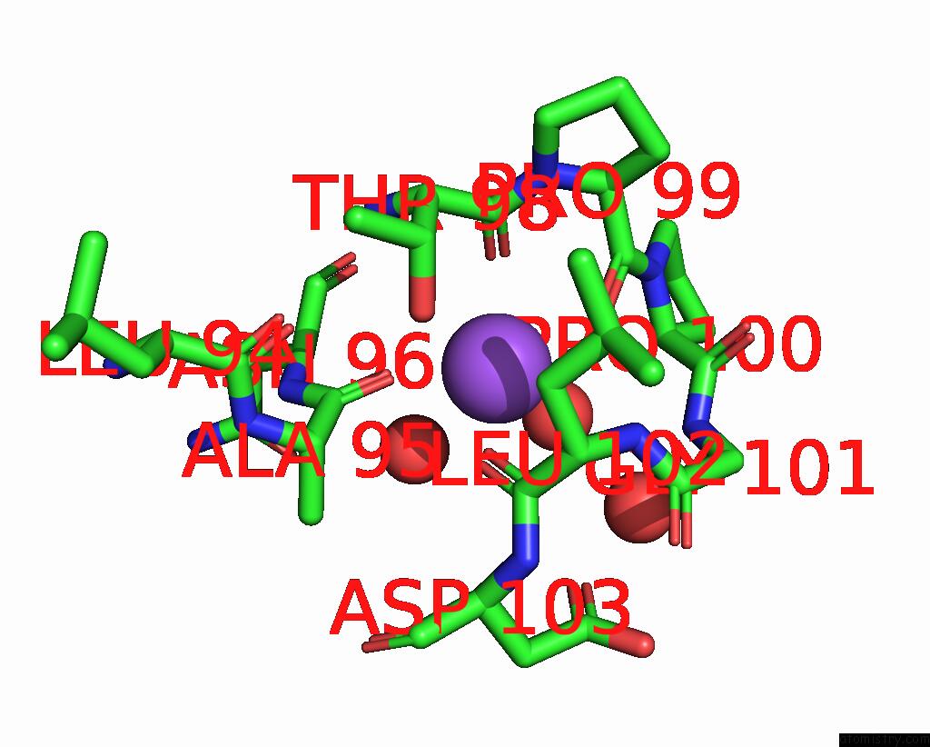
Mono view
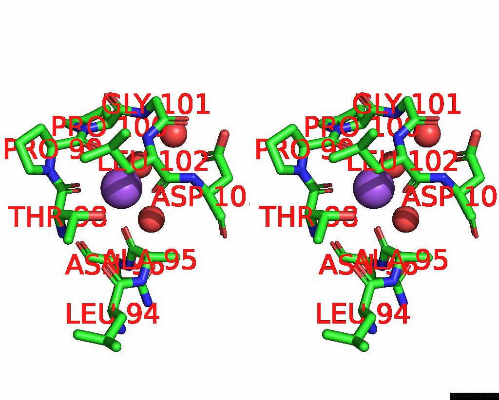
Stereo pair view
Mono view
Stereo pair view
A full contact list of Sodium with other atoms in the Na binding
site number 1 of Crystal Structure of Dialkylglycine Decarboxylases Bound with Calcium Ion within 5.0Å range:
|
Reference:
W.Liu,
M.D.Toney.
Crystal Structures of Dialkylglycine Decarboxylase Bound with Cesium Ion and Calcium Ion To Be Published.
Page generated: Mon Oct 7 01:43:26 2024
Last articles
Ca in 2WOBCa in 2WO4
Ca in 2WN3
Ca in 2WNX
Ca in 2WN2
Ca in 2WNV
Ca in 2WNP
Ca in 2WNO
Ca in 2WND
Ca in 2WM4