Sodium »
PDB 1z2u-1zud »
1z8j »
Sodium in PDB 1z8j: Crystal Structure of the Thrombin Mutant G193P Bound to Ppack
Enzymatic activity of Crystal Structure of the Thrombin Mutant G193P Bound to Ppack
All present enzymatic activity of Crystal Structure of the Thrombin Mutant G193P Bound to Ppack:
3.4.21.5;
3.4.21.5;
Protein crystallography data
The structure of Crystal Structure of the Thrombin Mutant G193P Bound to Ppack, PDB code: 1z8j
was solved by
K.M.Bobofchak,
A.O.Pineda,
F.S.Mathews,
E.Di Cera,
with X-Ray Crystallography technique. A brief refinement statistics is given in the table below:
| Resolution Low / High (Å) | 40.00 / 2.00 |
| Space group | P 21 21 21 |
| Cell size a, b, c (Å), α, β, γ (°) | 49.710, 73.550, 90.100, 90.00, 90.00, 90.00 |
| R / Rfree (%) | 20.5 / 24.6 |
Other elements in 1z8j:
The structure of Crystal Structure of the Thrombin Mutant G193P Bound to Ppack also contains other interesting chemical elements:
| Zinc | (Zn) | 3 atoms |
Sodium Binding Sites:
The binding sites of Sodium atom in the Crystal Structure of the Thrombin Mutant G193P Bound to Ppack
(pdb code 1z8j). This binding sites where shown within
5.0 Angstroms radius around Sodium atom.
In total only one binding site of Sodium was determined in the Crystal Structure of the Thrombin Mutant G193P Bound to Ppack, PDB code: 1z8j:
In total only one binding site of Sodium was determined in the Crystal Structure of the Thrombin Mutant G193P Bound to Ppack, PDB code: 1z8j:
Sodium binding site 1 out of 1 in 1z8j
Go back to
Sodium binding site 1 out
of 1 in the Crystal Structure of the Thrombin Mutant G193P Bound to Ppack
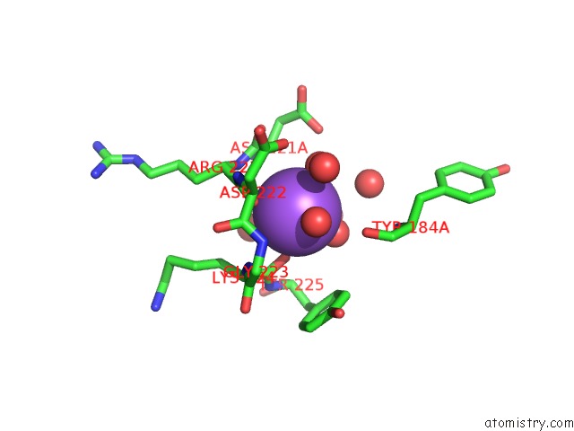
Mono view
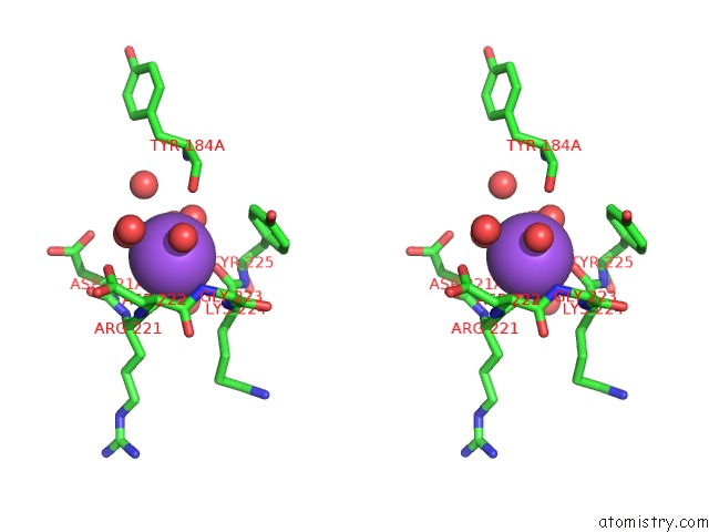
Stereo pair view
Mono view
Stereo pair view
A full contact list of Sodium with other atoms in the Na binding
site number 1 of Crystal Structure of the Thrombin Mutant G193P Bound to Ppack within 5.0Å range:
|
Reference:
K.M.Bobofchak,
A.O.Pineda,
F.S.Mathews,
E.Di Cera.
Energetic and Structural Consequences of Perturbing Gly-193 in the Oxyanion Hole of Serine Proteases J.Biol.Chem. V. 280 25644 2005.
ISSN: ISSN 0021-9258
PubMed: 15890651
DOI: 10.1074/JBC.M503499200
Page generated: Mon Oct 7 01:40:50 2024
ISSN: ISSN 0021-9258
PubMed: 15890651
DOI: 10.1074/JBC.M503499200
Last articles
Zn in 9J0NZn in 9J0O
Zn in 9J0P
Zn in 9FJX
Zn in 9EKB
Zn in 9C0F
Zn in 9CAH
Zn in 9CH0
Zn in 9CH3
Zn in 9CH1