Sodium »
PDB 1w5n-1x7d »
1wn2 »
Sodium in PDB 1wn2: Crystal Structure of Project Id PH1539 From Pyrococcus Horikoshii OT3
Enzymatic activity of Crystal Structure of Project Id PH1539 From Pyrococcus Horikoshii OT3
All present enzymatic activity of Crystal Structure of Project Id PH1539 From Pyrococcus Horikoshii OT3:
3.1.1.29;
3.1.1.29;
Protein crystallography data
The structure of Crystal Structure of Project Id PH1539 From Pyrococcus Horikoshii OT3, PDB code: 1wn2
was solved by
K.Shimizu,
N.Kunishima,
Riken Structural Genomics/Proteomics Initiative(Rsgi),
with X-Ray Crystallography technique. A brief refinement statistics is given in the table below:
| Resolution Low / High (Å) | 35.12 / 1.20 |
| Space group | P 41 21 2 |
| Cell size a, b, c (Å), α, β, γ (°) | 49.670, 49.670, 85.791, 90.00, 90.00, 90.00 |
| R / Rfree (%) | 19.3 / 21.5 |
Sodium Binding Sites:
The binding sites of Sodium atom in the Crystal Structure of Project Id PH1539 From Pyrococcus Horikoshii OT3
(pdb code 1wn2). This binding sites where shown within
5.0 Angstroms radius around Sodium atom.
In total only one binding site of Sodium was determined in the Crystal Structure of Project Id PH1539 From Pyrococcus Horikoshii OT3, PDB code: 1wn2:
In total only one binding site of Sodium was determined in the Crystal Structure of Project Id PH1539 From Pyrococcus Horikoshii OT3, PDB code: 1wn2:
Sodium binding site 1 out of 1 in 1wn2
Go back to
Sodium binding site 1 out
of 1 in the Crystal Structure of Project Id PH1539 From Pyrococcus Horikoshii OT3
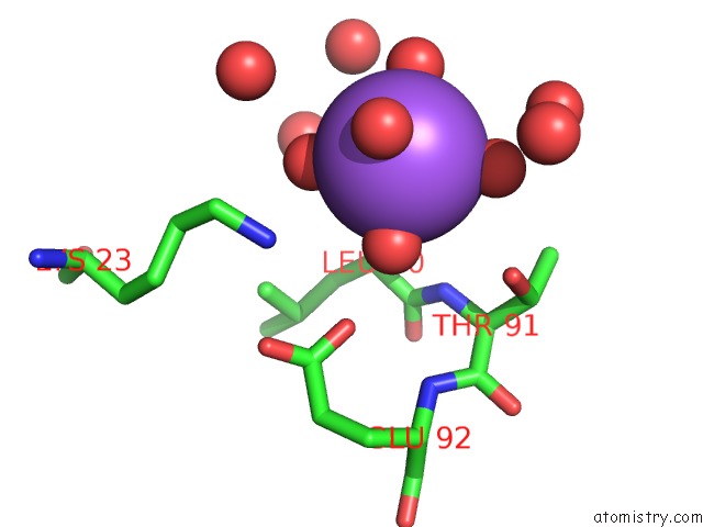
Mono view
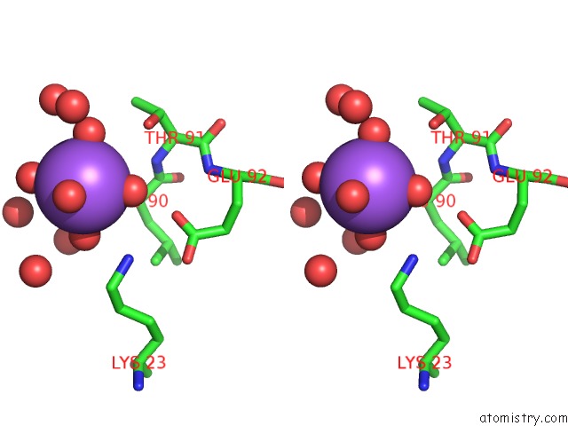
Stereo pair view
Mono view
Stereo pair view
A full contact list of Sodium with other atoms in the Na binding
site number 1 of Crystal Structure of Project Id PH1539 From Pyrococcus Horikoshii OT3 within 5.0Å range:
|
Reference:
K.Shimizu,
C.Kuroishi,
M.Sugahara,
N.Kunishima.
Structure of Peptidyl-Trna Hydrolase 2 From Pyrococcus Horikoshii OT3: Insight Into the Functional Role of Its Dimeric State. Acta Crystallogr.,Sect.D V. 64 444 2008.
ISSN: ISSN 0907-4449
PubMed: 18391411
DOI: 10.1107/S0907444908002850
Page generated: Mon Oct 7 00:26:19 2024
ISSN: ISSN 0907-4449
PubMed: 18391411
DOI: 10.1107/S0907444908002850
Last articles
Cl in 7WMTCl in 7WMK
Cl in 7WMI
Cl in 7WM7
Cl in 7WLW
Cl in 7WL9
Cl in 7WKZ
Cl in 7WKR
Cl in 7WGP
Cl in 7WK1