Sodium »
PDB 1ubs-1v54 »
1v47 »
Sodium in PDB 1v47: Crystal Structure of Atp Sulfurylase From Thermus Thermophillus HB8 in Complex with Aps
Enzymatic activity of Crystal Structure of Atp Sulfurylase From Thermus Thermophillus HB8 in Complex with Aps
All present enzymatic activity of Crystal Structure of Atp Sulfurylase From Thermus Thermophillus HB8 in Complex with Aps:
2.7.7.4;
2.7.7.4;
Protein crystallography data
The structure of Crystal Structure of Atp Sulfurylase From Thermus Thermophillus HB8 in Complex with Aps, PDB code: 1v47
was solved by
Y.Taguchi,
M.Sugishima,
K.Fukuyama,
Riken Structuralgenomics/Proteomics Initiative (Rsgi),
with X-Ray Crystallography technique. A brief refinement statistics is given in the table below:
| Resolution Low / High (Å) | 20.00 / 2.49 |
| Space group | P 1 21 1 |
| Cell size a, b, c (Å), α, β, γ (°) | 68.858, 61.254, 128.665, 90.00, 95.43, 90.00 |
| R / Rfree (%) | 21.9 / 26.9 |
Other elements in 1v47:
The structure of Crystal Structure of Atp Sulfurylase From Thermus Thermophillus HB8 in Complex with Aps also contains other interesting chemical elements:
| Zinc | (Zn) | 2 atoms |
| Chlorine | (Cl) | 5 atoms |
Sodium Binding Sites:
The binding sites of Sodium atom in the Crystal Structure of Atp Sulfurylase From Thermus Thermophillus HB8 in Complex with Aps
(pdb code 1v47). This binding sites where shown within
5.0 Angstroms radius around Sodium atom.
In total 4 binding sites of Sodium where determined in the Crystal Structure of Atp Sulfurylase From Thermus Thermophillus HB8 in Complex with Aps, PDB code: 1v47:
Jump to Sodium binding site number: 1; 2; 3; 4;
In total 4 binding sites of Sodium where determined in the Crystal Structure of Atp Sulfurylase From Thermus Thermophillus HB8 in Complex with Aps, PDB code: 1v47:
Jump to Sodium binding site number: 1; 2; 3; 4;
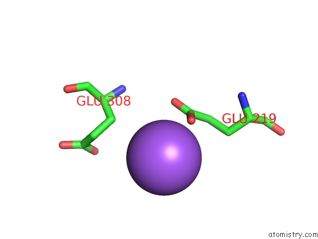
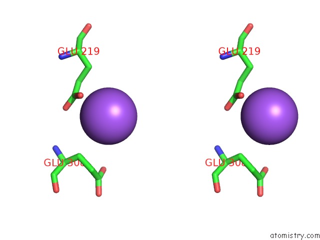
Sodium binding site 1 out of 4 in 1v47
Go back to
Sodium binding site 1 out
of 4 in the Crystal Structure of Atp Sulfurylase From Thermus Thermophillus HB8 in Complex with Aps
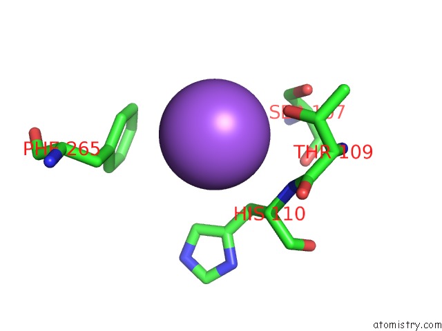
Mono view
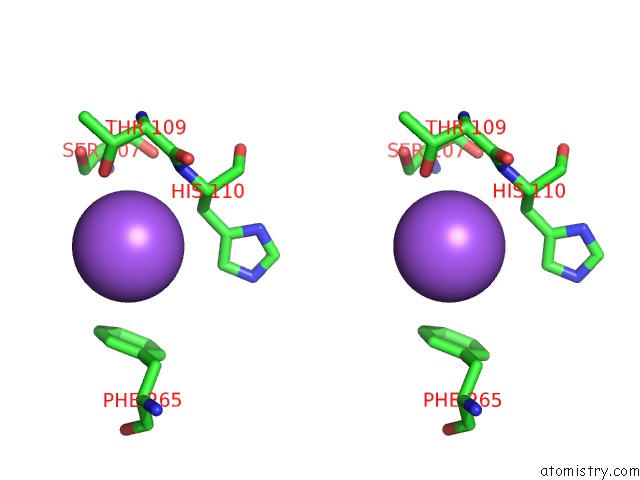
Stereo pair view
Mono view
Stereo pair view
A full contact list of Sodium with other atoms in the Na binding
site number 1 of Crystal Structure of Atp Sulfurylase From Thermus Thermophillus HB8 in Complex with Aps within 5.0Å range:
|
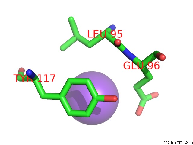
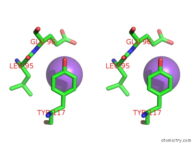
Sodium binding site 2 out of 4 in 1v47
Go back to
Sodium binding site 2 out
of 4 in the Crystal Structure of Atp Sulfurylase From Thermus Thermophillus HB8 in Complex with Aps
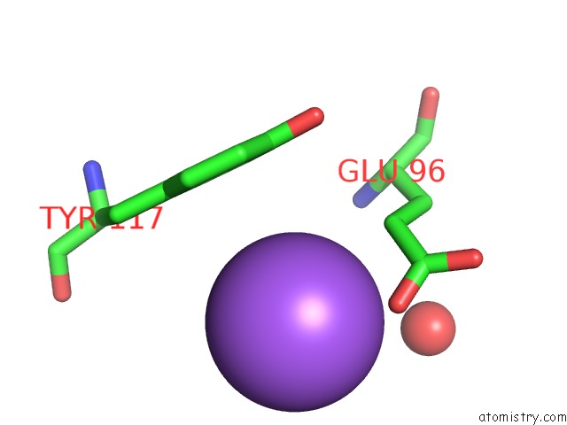
Mono view
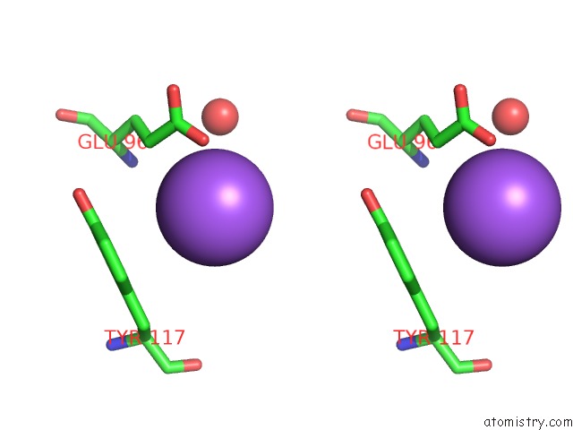
Stereo pair view
Mono view
Stereo pair view
A full contact list of Sodium with other atoms in the Na binding
site number 2 of Crystal Structure of Atp Sulfurylase From Thermus Thermophillus HB8 in Complex with Aps within 5.0Å range:
|
Sodium binding site 3 out of 4 in 1v47
Go back to
Sodium binding site 3 out
of 4 in the Crystal Structure of Atp Sulfurylase From Thermus Thermophillus HB8 in Complex with Aps
Mono view
Stereo pair view
Mono view
Stereo pair view
A full contact list of Sodium with other atoms in the Na binding
site number 3 of Crystal Structure of Atp Sulfurylase From Thermus Thermophillus HB8 in Complex with Aps within 5.0Å range:
|
Sodium binding site 4 out of 4 in 1v47
Go back to
Sodium binding site 4 out
of 4 in the Crystal Structure of Atp Sulfurylase From Thermus Thermophillus HB8 in Complex with Aps
Mono view
Stereo pair view
Mono view
Stereo pair view
A full contact list of Sodium with other atoms in the Na binding
site number 4 of Crystal Structure of Atp Sulfurylase From Thermus Thermophillus HB8 in Complex with Aps within 5.0Å range:
|
Reference:
Y.Taguchi,
M.Sugishima,
K.Fukuyama.
Crystal Structure of A Novel Zinc-Binding Atp Sulfurylase From Thermus Thermophilus HB8 Biochemistry V. 43 4111 2004.
ISSN: ISSN 0006-2960
PubMed: 15065853
DOI: 10.1021/BI036052T
Page generated: Sun Oct 6 22:49:30 2024
ISSN: ISSN 0006-2960
PubMed: 15065853
DOI: 10.1021/BI036052T
Last articles
Cl in 5QCHCl in 5QD5
Cl in 5QE6
Cl in 5QCN
Cl in 5QCM
Cl in 5QCG
Cl in 5QCL
Cl in 5QCK
Cl in 5QCE
Cl in 5QCI