Sodium »
PDB 1t4b-1u8r »
1txz »
Sodium in PDB 1txz: Crystal Structure of Yeast YMX7, An Adp-Ribose-1''-Monophosphatase, Complexed with Adp-Ribose
Protein crystallography data
The structure of Crystal Structure of Yeast YMX7, An Adp-Ribose-1''-Monophosphatase, Complexed with Adp-Ribose, PDB code: 1txz
was solved by
D.Kumaran,
S.Swaminathan,
S.K.Burley,
New York Sgx Research Center Forstructural Genomics (Nysgxrc),
with X-Ray Crystallography technique. A brief refinement statistics is given in the table below:
| Resolution Low / High (Å) | 50.00 / 2.05 |
| Space group | C 1 2 1 |
| Cell size a, b, c (Å), α, β, γ (°) | 107.437, 37.931, 62.514, 90.00, 112.30, 90.00 |
| R / Rfree (%) | 20 / 23.5 |
Sodium Binding Sites:
The binding sites of Sodium atom in the Crystal Structure of Yeast YMX7, An Adp-Ribose-1''-Monophosphatase, Complexed with Adp-Ribose
(pdb code 1txz). This binding sites where shown within
5.0 Angstroms radius around Sodium atom.
In total only one binding site of Sodium was determined in the Crystal Structure of Yeast YMX7, An Adp-Ribose-1''-Monophosphatase, Complexed with Adp-Ribose, PDB code: 1txz:
In total only one binding site of Sodium was determined in the Crystal Structure of Yeast YMX7, An Adp-Ribose-1''-Monophosphatase, Complexed with Adp-Ribose, PDB code: 1txz:
Sodium binding site 1 out of 1 in 1txz
Go back to
Sodium binding site 1 out
of 1 in the Crystal Structure of Yeast YMX7, An Adp-Ribose-1''-Monophosphatase, Complexed with Adp-Ribose
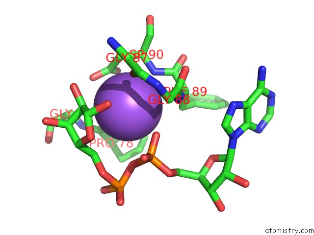
Mono view
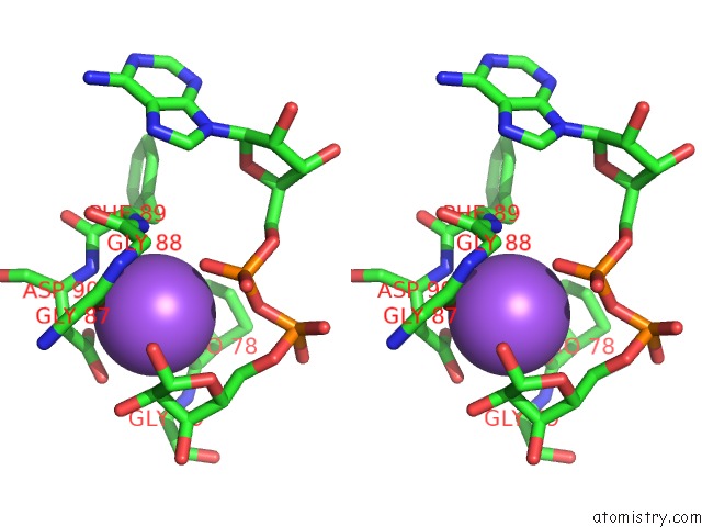
Stereo pair view
Mono view
Stereo pair view
A full contact list of Sodium with other atoms in the Na binding
site number 1 of Crystal Structure of Yeast YMX7, An Adp-Ribose-1''-Monophosphatase, Complexed with Adp-Ribose within 5.0Å range:
|
Reference:
D.Kumaran,
S.Eswaramoorthy,
F.W.Studier,
S.Swaminathan.
Structure and Mechanism of Adp-Ribose-1''-Monophosphatase (Appr-1''-Pase), A Ubiquitous Cellular Processing Enzyme. Protein Sci. V. 14 719 2005.
ISSN: ISSN 0961-8368
PubMed: 15722447
DOI: 10.1110/PS.041132005
Page generated: Sun Oct 6 22:42:10 2024
ISSN: ISSN 0961-8368
PubMed: 15722447
DOI: 10.1110/PS.041132005
Last articles
Zn in 9MJ5Zn in 9HNW
Zn in 9G0L
Zn in 9FNE
Zn in 9DZN
Zn in 9E0I
Zn in 9D32
Zn in 9DAK
Zn in 8ZXC
Zn in 8ZUF