Sodium »
PDB 1t4b-1u8r »
1tm1 »
Sodium in PDB 1tm1: Crystal Structure of the Complex of Subtilisin Bpn' with Chymotrypsin Inhibitor 2
Enzymatic activity of Crystal Structure of the Complex of Subtilisin Bpn' with Chymotrypsin Inhibitor 2
All present enzymatic activity of Crystal Structure of the Complex of Subtilisin Bpn' with Chymotrypsin Inhibitor 2:
3.4.21.62;
3.4.21.62;
Protein crystallography data
The structure of Crystal Structure of the Complex of Subtilisin Bpn' with Chymotrypsin Inhibitor 2, PDB code: 1tm1
was solved by
E.S.Radisky,
G.Kwan,
C.J.Karen Lu,
D.E.Koshland Jr.,
with X-Ray Crystallography technique. A brief refinement statistics is given in the table below:
| Resolution Low / High (Å) | 81.65 / 1.70 |
| Space group | P 65 2 2 |
| Cell size a, b, c (Å), α, β, γ (°) | 93.720, 93.720, 185.853, 90.00, 90.00, 120.00 |
| R / Rfree (%) | 15.1 / 18.2 |
Other elements in 1tm1:
The structure of Crystal Structure of the Complex of Subtilisin Bpn' with Chymotrypsin Inhibitor 2 also contains other interesting chemical elements:
| Calcium | (Ca) | 1 atom |
Sodium Binding Sites:
The binding sites of Sodium atom in the Crystal Structure of the Complex of Subtilisin Bpn' with Chymotrypsin Inhibitor 2
(pdb code 1tm1). This binding sites where shown within
5.0 Angstroms radius around Sodium atom.
In total only one binding site of Sodium was determined in the Crystal Structure of the Complex of Subtilisin Bpn' with Chymotrypsin Inhibitor 2, PDB code: 1tm1:
In total only one binding site of Sodium was determined in the Crystal Structure of the Complex of Subtilisin Bpn' with Chymotrypsin Inhibitor 2, PDB code: 1tm1:
Sodium binding site 1 out of 1 in 1tm1
Go back to
Sodium binding site 1 out
of 1 in the Crystal Structure of the Complex of Subtilisin Bpn' with Chymotrypsin Inhibitor 2
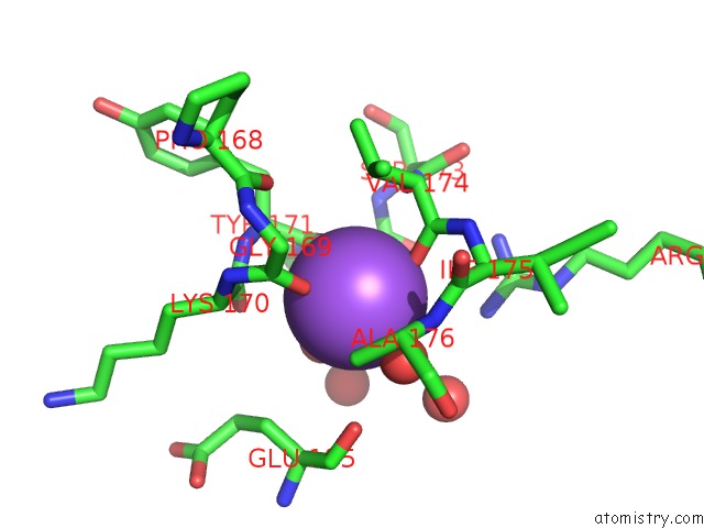
Mono view
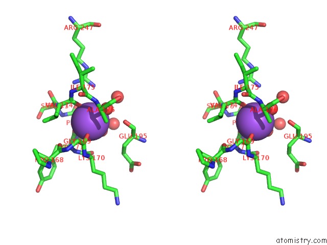
Stereo pair view
Mono view
Stereo pair view
A full contact list of Sodium with other atoms in the Na binding
site number 1 of Crystal Structure of the Complex of Subtilisin Bpn' with Chymotrypsin Inhibitor 2 within 5.0Å range:
|
Reference:
E.S.Radisky,
G.Kwan,
C.J.Karen Lu,
D.E.Koshland Jr..
Binding, Proteolytic, and Crystallographic Analyses of Mutations at the Protease-Inhibitor Interface of the Subtilisin Bpn'/Chymotrypsin Inhibitor 2 Complex(,). Biochemistry V. 43 13648 2004.
ISSN: ISSN 0006-2960
PubMed: 15504027
DOI: 10.1021/BI048797K
Page generated: Sun Oct 6 22:40:25 2024
ISSN: ISSN 0006-2960
PubMed: 15504027
DOI: 10.1021/BI048797K
Last articles
Cl in 5G5NCl in 5G5O
Cl in 5G5G
Cl in 5G60
Cl in 5G5H
Cl in 5G3T
Cl in 5G54
Cl in 5G4A
Cl in 5G4Q
Cl in 5G47