Sodium »
PDB 1oar-1ph6 »
1oco »
Sodium in PDB 1oco: Bovine Heart Cytochrome C Oxidase in Carbon Monoxide-Bound State
Enzymatic activity of Bovine Heart Cytochrome C Oxidase in Carbon Monoxide-Bound State
All present enzymatic activity of Bovine Heart Cytochrome C Oxidase in Carbon Monoxide-Bound State:
1.9.3.1;
1.9.3.1;
Protein crystallography data
The structure of Bovine Heart Cytochrome C Oxidase in Carbon Monoxide-Bound State, PDB code: 1oco
was solved by
T.Tsukihara,
M.Yao,
with X-Ray Crystallography technique. A brief refinement statistics is given in the table below:
| Resolution Low / High (Å) | 7.00 / 2.80 |
| Space group | P 21 21 21 |
| Cell size a, b, c (Å), α, β, γ (°) | 189.100, 210.500, 178.600, 90.00, 90.00, 90.00 |
| R / Rfree (%) | 21.3 / 25.6 |
Other elements in 1oco:
The structure of Bovine Heart Cytochrome C Oxidase in Carbon Monoxide-Bound State also contains other interesting chemical elements:
| Magnesium | (Mg) | 2 atoms |
| Zinc | (Zn) | 2 atoms |
| Iron | (Fe) | 4 atoms |
| Copper | (Cu) | 6 atoms |
Sodium Binding Sites:
The binding sites of Sodium atom in the Bovine Heart Cytochrome C Oxidase in Carbon Monoxide-Bound State
(pdb code 1oco). This binding sites where shown within
5.0 Angstroms radius around Sodium atom.
In total 2 binding sites of Sodium where determined in the Bovine Heart Cytochrome C Oxidase in Carbon Monoxide-Bound State, PDB code: 1oco:
Jump to Sodium binding site number: 1; 2;
In total 2 binding sites of Sodium where determined in the Bovine Heart Cytochrome C Oxidase in Carbon Monoxide-Bound State, PDB code: 1oco:
Jump to Sodium binding site number: 1; 2;
Sodium binding site 1 out of 2 in 1oco
Go back to
Sodium binding site 1 out
of 2 in the Bovine Heart Cytochrome C Oxidase in Carbon Monoxide-Bound State
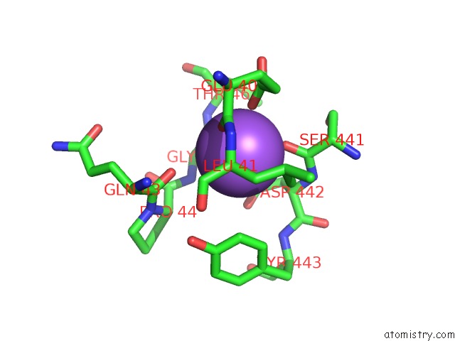
Mono view
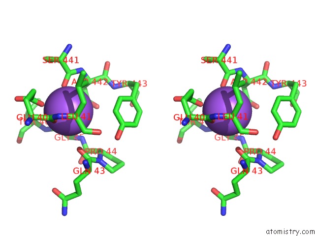
Stereo pair view
Mono view
Stereo pair view
A full contact list of Sodium with other atoms in the Na binding
site number 1 of Bovine Heart Cytochrome C Oxidase in Carbon Monoxide-Bound State within 5.0Å range:
|
Sodium binding site 2 out of 2 in 1oco
Go back to
Sodium binding site 2 out
of 2 in the Bovine Heart Cytochrome C Oxidase in Carbon Monoxide-Bound State
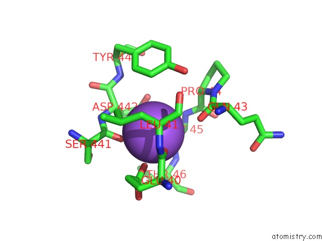
Mono view
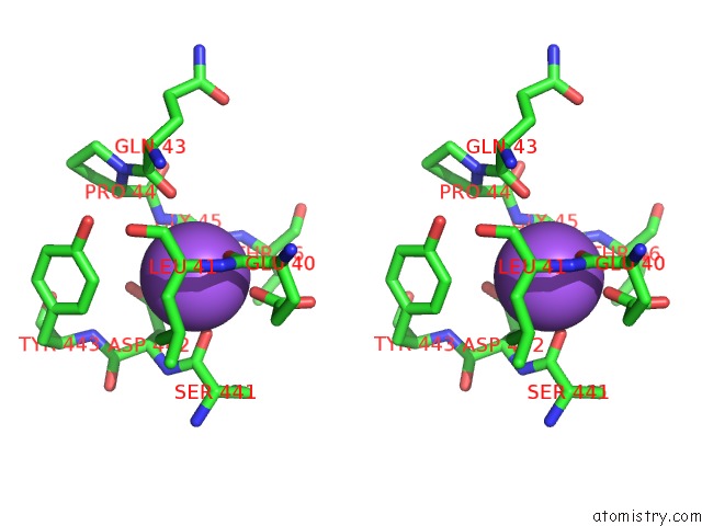
Stereo pair view
Mono view
Stereo pair view
A full contact list of Sodium with other atoms in the Na binding
site number 2 of Bovine Heart Cytochrome C Oxidase in Carbon Monoxide-Bound State within 5.0Å range:
|
Reference:
S.Yoshikawa,
K.Shinzawa-Itoh,
R.Nakashima,
R.Yaono,
E.Yamashita,
N.Inoue,
M.Yao,
M.J.Fei,
C.P.Libeu,
T.Mizushima,
H.Yamaguchi,
T.Tomizaki,
T.Tsukihara.
Redox-Coupled Crystal Structural Changes in Bovine Heart Cytochrome C Oxidase. Science V. 280 1723 1998.
ISSN: ISSN 0036-8075
PubMed: 9624044
DOI: 10.1126/SCIENCE.280.5370.1723
Page generated: Sun Oct 6 21:11:06 2024
ISSN: ISSN 0036-8075
PubMed: 9624044
DOI: 10.1126/SCIENCE.280.5370.1723
Last articles
Fe in 2YXOFe in 2YRS
Fe in 2YXC
Fe in 2YNM
Fe in 2YVJ
Fe in 2YP1
Fe in 2YU2
Fe in 2YU1
Fe in 2YQB
Fe in 2YOO