Sodium »
PDB 1oar-1ph6 »
1ob7 »
Sodium in PDB 1ob7: Cephaibol C
Protein crystallography data
The structure of Cephaibol C, PDB code: 1ob7
was solved by
G.Bunkoczi,
M.Schiell,
L.Vertesy,
G.M.Sheldrick,
with X-Ray Crystallography technique. A brief refinement statistics is given in the table below:
| Resolution Low / High (Å) | 28.62 / 0.89 |
| Space group | P 21 21 21 |
| Cell size a, b, c (Å), α, β, γ (°) | 9.002, 28.619, 41.100, 90.00, 90.00, 90.00 |
| R / Rfree (%) | 7.2 / 7.7 |
Sodium Binding Sites:
The binding sites of Sodium atom in the Cephaibol C
(pdb code 1ob7). This binding sites where shown within
5.0 Angstroms radius around Sodium atom.
In total only one binding site of Sodium was determined in the Cephaibol C, PDB code: 1ob7:
In total only one binding site of Sodium was determined in the Cephaibol C, PDB code: 1ob7:
Sodium binding site 1 out of 1 in 1ob7
Go back to
Sodium binding site 1 out
of 1 in the Cephaibol C

Mono view
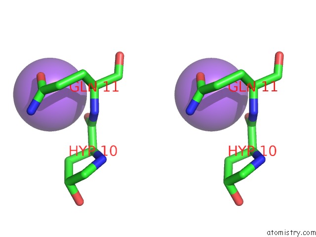
Stereo pair view
Mono view
Stereo pair view
A full contact list of Sodium with other atoms in the Na binding
site number 1 of Cephaibol C within 5.0Å range:
|
Reference:
G.Bunkoczi,
M.Schiell,
L.Vertesy,
G.M.Sheldrick.
Crystal Structures of Cephaibols J.Pept.Sci. V. 9 745 2003.
ISSN: ISSN 1075-2617
PubMed: 14658793
DOI: 10.1002/PSC.496
Page generated: Sun Aug 17 06:52:04 2025
ISSN: ISSN 1075-2617
PubMed: 14658793
DOI: 10.1002/PSC.496
Last articles
Na in 4Y90Na in 4Y9V
Na in 4Y96
Na in 4XTN
Na in 4Y8F
Na in 4Y7D
Na in 4Y0W
Na in 4Y6G
Na in 4Y5L
Na in 4Y23