Sodium »
PDB 1m90-1nji »
1mmu »
Sodium in PDB 1mmu: Crystal Structure of Galactose Mutarotase From Lactococcus Lactis Complexed with D-Glucose
Enzymatic activity of Crystal Structure of Galactose Mutarotase From Lactococcus Lactis Complexed with D-Glucose
All present enzymatic activity of Crystal Structure of Galactose Mutarotase From Lactococcus Lactis Complexed with D-Glucose:
5.1.3.3;
5.1.3.3;
Protein crystallography data
The structure of Crystal Structure of Galactose Mutarotase From Lactococcus Lactis Complexed with D-Glucose, PDB code: 1mmu
was solved by
J.B.Thoden,
J.Kim,
F.M.Raushel,
H.M.Holden,
with X-Ray Crystallography technique. A brief refinement statistics is given in the table below:
| Resolution Low / High (Å) | 30.00 / 1.80 |
| Space group | P 21 21 21 |
| Cell size a, b, c (Å), α, β, γ (°) | 44.200, 75.600, 209.500, 90.00, 90.00, 90.00 |
| R / Rfree (%) | n/a / n/a |
Sodium Binding Sites:
The binding sites of Sodium atom in the Crystal Structure of Galactose Mutarotase From Lactococcus Lactis Complexed with D-Glucose
(pdb code 1mmu). This binding sites where shown within
5.0 Angstroms radius around Sodium atom.
In total only one binding site of Sodium was determined in the Crystal Structure of Galactose Mutarotase From Lactococcus Lactis Complexed with D-Glucose, PDB code: 1mmu:
In total only one binding site of Sodium was determined in the Crystal Structure of Galactose Mutarotase From Lactococcus Lactis Complexed with D-Glucose, PDB code: 1mmu:
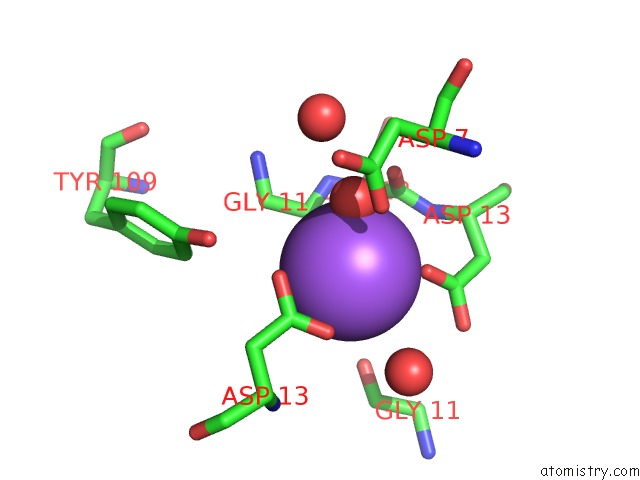
Sodium binding site 1 out of 1 in 1mmu
Go back to
Sodium binding site 1 out
of 1 in the Crystal Structure of Galactose Mutarotase From Lactococcus Lactis Complexed with D-Glucose
Mono view

Stereo pair view
Mono view
Stereo pair view
A full contact list of Sodium with other atoms in the Na binding
site number 1 of Crystal Structure of Galactose Mutarotase From Lactococcus Lactis Complexed with D-Glucose within 5.0Å range:
|
Reference:
J.B.Thoden,
J.Kim,
F.M.Raushel,
H.M.Holden.
Structural and Kinetic Studies of Sugar Binding to Galactose Mutarotase From Lactococcus Lactis. J.Biol.Chem. V. 277 45458 2002.
ISSN: ISSN 0021-9258
PubMed: 12218067
DOI: 10.1074/JBC.M208395200
Page generated: Sun Oct 6 20:32:33 2024
ISSN: ISSN 0021-9258
PubMed: 12218067
DOI: 10.1074/JBC.M208395200
Last articles
Zn in 9MJ5Zn in 9HNW
Zn in 9G0L
Zn in 9FNE
Zn in 9DZN
Zn in 9E0I
Zn in 9D32
Zn in 9DAK
Zn in 8ZXC
Zn in 8ZUF