Sodium »
PDB 1m90-1nji »
1me8 »
Sodium in PDB 1me8: Inosine Monophosphate Dehydrogenase (Impdh) From Tritrichomonas Foetus with Rvp Bound
Enzymatic activity of Inosine Monophosphate Dehydrogenase (Impdh) From Tritrichomonas Foetus with Rvp Bound
All present enzymatic activity of Inosine Monophosphate Dehydrogenase (Impdh) From Tritrichomonas Foetus with Rvp Bound:
1.1.1.205;
1.1.1.205;
Protein crystallography data
The structure of Inosine Monophosphate Dehydrogenase (Impdh) From Tritrichomonas Foetus with Rvp Bound, PDB code: 1me8
was solved by
G.L.Prosise,
J.Wu,
H.Luecke,
with X-Ray Crystallography technique. A brief refinement statistics is given in the table below:
| Resolution Low / High (Å) | 48.94 / 1.90 |
| Space group | P 4 3 2 |
| Cell size a, b, c (Å), α, β, γ (°) | 154.745, 154.745, 154.745, 90.00, 90.00, 90.00 |
| R / Rfree (%) | 24.3 / 25.8 |
Other elements in 1me8:
The structure of Inosine Monophosphate Dehydrogenase (Impdh) From Tritrichomonas Foetus with Rvp Bound also contains other interesting chemical elements:
| Potassium | (K) | 1 atom |
Sodium Binding Sites:
The binding sites of Sodium atom in the Inosine Monophosphate Dehydrogenase (Impdh) From Tritrichomonas Foetus with Rvp Bound
(pdb code 1me8). This binding sites where shown within
5.0 Angstroms radius around Sodium atom.
In total only one binding site of Sodium was determined in the Inosine Monophosphate Dehydrogenase (Impdh) From Tritrichomonas Foetus with Rvp Bound, PDB code: 1me8:
In total only one binding site of Sodium was determined in the Inosine Monophosphate Dehydrogenase (Impdh) From Tritrichomonas Foetus with Rvp Bound, PDB code: 1me8:
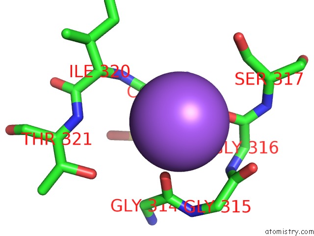
Sodium binding site 1 out of 1 in 1me8
Go back to
Sodium binding site 1 out
of 1 in the Inosine Monophosphate Dehydrogenase (Impdh) From Tritrichomonas Foetus with Rvp Bound
Mono view
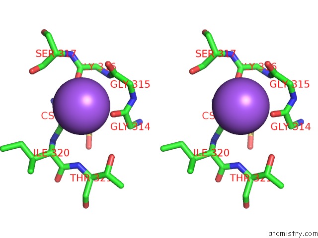
Stereo pair view
Mono view
Stereo pair view
A full contact list of Sodium with other atoms in the Na binding
site number 1 of Inosine Monophosphate Dehydrogenase (Impdh) From Tritrichomonas Foetus with Rvp Bound within 5.0Å range:
|
Reference:
G.L.Prosise,
J.Wu,
H.Luecke.
Crystal Structure of Tritrichomonas Foetus Inosine Monophosphate Dehydrogenase in Complex with the Inhibitor Ribavirin Monophosphate Reveals A Catalysis-Dependent Ion-Binding Site J.Biol.Chem. V. 277 50654 2002.
ISSN: ISSN 0021-9258
PubMed: 12235158
DOI: 10.1074/JBC.M208330200
Page generated: Sun Oct 6 20:31:31 2024
ISSN: ISSN 0021-9258
PubMed: 12235158
DOI: 10.1074/JBC.M208330200
Last articles
Zn in 9MJ5Zn in 9HNW
Zn in 9G0L
Zn in 9FNE
Zn in 9DZN
Zn in 9E0I
Zn in 9D32
Zn in 9DAK
Zn in 8ZXC
Zn in 8ZUF