Sodium »
PDB 1l0r-1m65 »
1l5b »
Sodium in PDB 1l5b: Domain-Swapped Cyanovirin-N Dimer
Protein crystallography data
The structure of Domain-Swapped Cyanovirin-N Dimer, PDB code: 1l5b
was solved by
L.G.Barrientos,
J.M.Louis,
I.Botos,
T.Mori,
Z.Han,
B.R.O'keefe,
M.R.Boyd,
A.Wlodawer,
A.M.Gronenborn,
with X-Ray Crystallography technique. A brief refinement statistics is given in the table below:
| Resolution Low / High (Å) | 19.90 / 2.00 |
| Space group | P 41 21 2 |
| Cell size a, b, c (Å), α, β, γ (°) | 61.966, 61.966, 148.400, 90.00, 90.00, 90.00 |
| R / Rfree (%) | 24.7 / 25.8 |
Sodium Binding Sites:
The binding sites of Sodium atom in the Domain-Swapped Cyanovirin-N Dimer
(pdb code 1l5b). This binding sites where shown within
5.0 Angstroms radius around Sodium atom.
In total only one binding site of Sodium was determined in the Domain-Swapped Cyanovirin-N Dimer, PDB code: 1l5b:
In total only one binding site of Sodium was determined in the Domain-Swapped Cyanovirin-N Dimer, PDB code: 1l5b:
Sodium binding site 1 out of 1 in 1l5b
Go back to
Sodium binding site 1 out
of 1 in the Domain-Swapped Cyanovirin-N Dimer
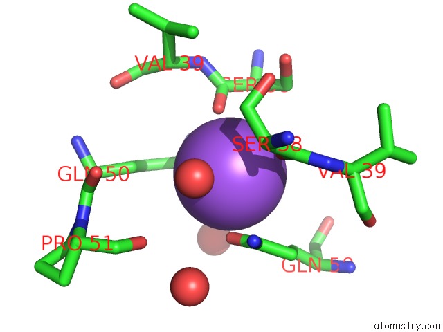
Mono view
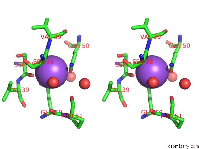
Stereo pair view
Mono view
Stereo pair view
A full contact list of Sodium with other atoms in the Na binding
site number 1 of Domain-Swapped Cyanovirin-N Dimer within 5.0Å range:
|
Reference:
L.G.Barrientos,
J.M.Louis,
I.Botos,
T.Mori,
Z.Han,
B.R.O'keefe,
M.R.Boyd,
A.Wlodawer,
A.M.Gronenborn.
The Domain-Swapped Dimer of Cyanovirin-N Is in A Metastable Folded State: Reconciliation of X-Ray and uc(Nmr) Structures. Structure V. 10 673 2002.
ISSN: ISSN 0969-2126
PubMed: 12015150
DOI: 10.1016/S0969-2126(02)00758-X
Page generated: Sun Oct 6 20:15:41 2024
ISSN: ISSN 0969-2126
PubMed: 12015150
DOI: 10.1016/S0969-2126(02)00758-X
Last articles
F in 7QMAF in 7QM6
F in 7QM9
F in 7QM8
F in 7QM7
F in 7QM5
F in 7QM4
F in 7QLT
F in 7QIT
F in 7QM3