Sodium »
PDB 1evr-1gb5 »
1g1s »
Sodium in PDB 1g1s: P-Selectin Lectin/Egf Domains Complexed with Psgl-1 Peptide
Protein crystallography data
The structure of P-Selectin Lectin/Egf Domains Complexed with Psgl-1 Peptide, PDB code: 1g1s
was solved by
W.S.Somers,
R.T.Camphausen,
with X-Ray Crystallography technique. A brief refinement statistics is given in the table below:
| Resolution Low / High (Å) | 15.00 / 1.90 |
| Space group | I 2 2 2 |
| Cell size a, b, c (Å), α, β, γ (°) | 63.445, 96.756, 187.289, 90.00, 90.00, 90.00 |
| R / Rfree (%) | 20.4 / 23.5 |
Other elements in 1g1s:
The structure of P-Selectin Lectin/Egf Domains Complexed with Psgl-1 Peptide also contains other interesting chemical elements:
| Strontium | (Sr) | 2 atoms |
Sodium Binding Sites:
The binding sites of Sodium atom in the P-Selectin Lectin/Egf Domains Complexed with Psgl-1 Peptide
(pdb code 1g1s). This binding sites where shown within
5.0 Angstroms radius around Sodium atom.
In total 2 binding sites of Sodium where determined in the P-Selectin Lectin/Egf Domains Complexed with Psgl-1 Peptide, PDB code: 1g1s:
Jump to Sodium binding site number: 1; 2;
In total 2 binding sites of Sodium where determined in the P-Selectin Lectin/Egf Domains Complexed with Psgl-1 Peptide, PDB code: 1g1s:
Jump to Sodium binding site number: 1; 2;
Sodium binding site 1 out of 2 in 1g1s
Go back to
Sodium binding site 1 out
of 2 in the P-Selectin Lectin/Egf Domains Complexed with Psgl-1 Peptide
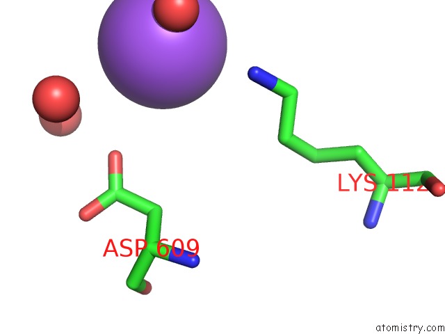
Mono view
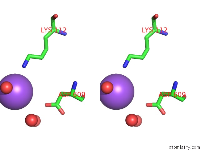
Stereo pair view
Mono view
Stereo pair view
A full contact list of Sodium with other atoms in the Na binding
site number 1 of P-Selectin Lectin/Egf Domains Complexed with Psgl-1 Peptide within 5.0Å range:
|
Sodium binding site 2 out of 2 in 1g1s
Go back to
Sodium binding site 2 out
of 2 in the P-Selectin Lectin/Egf Domains Complexed with Psgl-1 Peptide
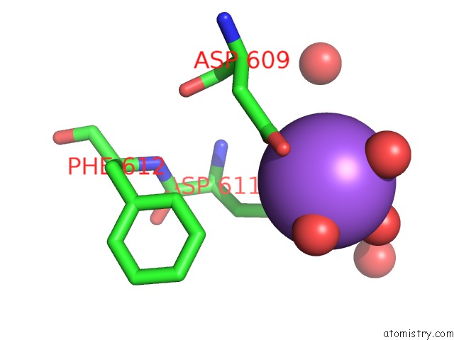
Mono view
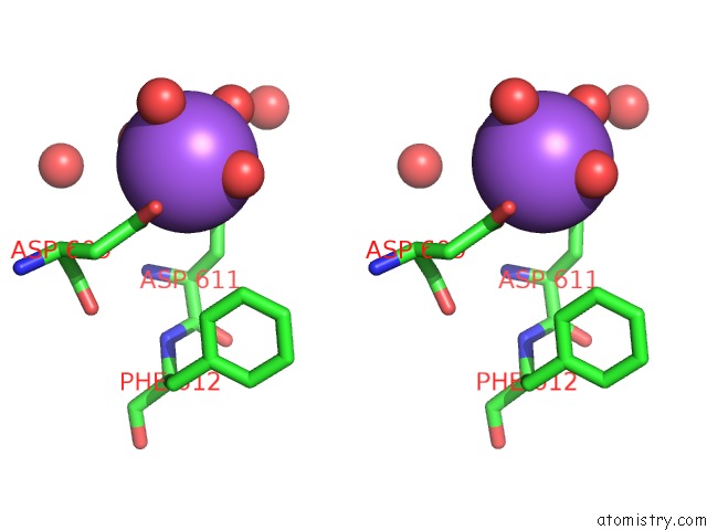
Stereo pair view
Mono view
Stereo pair view
A full contact list of Sodium with other atoms in the Na binding
site number 2 of P-Selectin Lectin/Egf Domains Complexed with Psgl-1 Peptide within 5.0Å range:
|
Reference:
W.S.Somers,
J.Tang,
G.D.Shaw,
R.T.Camphausen.
Insights Into the Molecular Basis of Leukocyte Tethering and Rolling Revealed By Structures of P- and E-Selectin Bound to Sle(X) and Psgl-1. Cell(Cambridge,Mass.) V. 103 467 2000.
ISSN: ISSN 0092-8674
PubMed: 11081633
DOI: 10.1016/S0092-8674(00)00138-0
Page generated: Sun Oct 6 18:29:12 2024
ISSN: ISSN 0092-8674
PubMed: 11081633
DOI: 10.1016/S0092-8674(00)00138-0
Last articles
Zn in 9J0NZn in 9J0O
Zn in 9J0P
Zn in 9FJX
Zn in 9EKB
Zn in 9C0F
Zn in 9CAH
Zn in 9CH0
Zn in 9CH3
Zn in 9CH1