Sodium »
PDB 1evr-1gb5 »
1f6o »
Sodium in PDB 1f6o: Crystal Structure of the Human Aag Dna Repair Glycosylase Complexed with Dna
Enzymatic activity of Crystal Structure of the Human Aag Dna Repair Glycosylase Complexed with Dna
All present enzymatic activity of Crystal Structure of the Human Aag Dna Repair Glycosylase Complexed with Dna:
3.2.2.20;
3.2.2.20;
Protein crystallography data
The structure of Crystal Structure of the Human Aag Dna Repair Glycosylase Complexed with Dna, PDB code: 1f6o
was solved by
A.Y.Lau,
M.D.Wyatt,
B.J.Glassner,
L.D.Samson,
T.Ellenberger,
with X-Ray Crystallography technique. A brief refinement statistics is given in the table below:
| Resolution Low / High (Å) | 500.00 / 2.40 |
| Space group | P 21 21 21 |
| Cell size a, b, c (Å), α, β, γ (°) | 43.750, 57.100, 128.450, 90.00, 90.00, 90.00 |
| R / Rfree (%) | 21.9 / 28.2 |
Sodium Binding Sites:
The binding sites of Sodium atom in the Crystal Structure of the Human Aag Dna Repair Glycosylase Complexed with Dna
(pdb code 1f6o). This binding sites where shown within
5.0 Angstroms radius around Sodium atom.
In total only one binding site of Sodium was determined in the Crystal Structure of the Human Aag Dna Repair Glycosylase Complexed with Dna, PDB code: 1f6o:
In total only one binding site of Sodium was determined in the Crystal Structure of the Human Aag Dna Repair Glycosylase Complexed with Dna, PDB code: 1f6o:
Sodium binding site 1 out of 1 in 1f6o
Go back to
Sodium binding site 1 out
of 1 in the Crystal Structure of the Human Aag Dna Repair Glycosylase Complexed with Dna
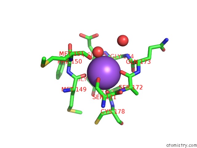
Mono view
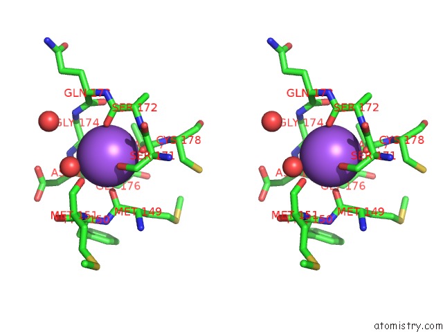
Stereo pair view
Mono view
Stereo pair view
A full contact list of Sodium with other atoms in the Na binding
site number 1 of Crystal Structure of the Human Aag Dna Repair Glycosylase Complexed with Dna within 5.0Å range:
|
Reference:
A.Y.Lau,
M.D.Wyatt,
B.J.Glassner,
L.D.Samson,
T.Ellenberger.
Molecular Basis For Discriminating Between Normal and Damaged Bases By the Human Alkyladenine Glycosylase, Aag. Proc.Natl.Acad.Sci.Usa V. 97 13573 2000.
ISSN: ISSN 0027-8424
PubMed: 11106395
DOI: 10.1073/PNAS.97.25.13573
Page generated: Sun Oct 6 18:28:02 2024
ISSN: ISSN 0027-8424
PubMed: 11106395
DOI: 10.1073/PNAS.97.25.13573
Last articles
Zn in 9J0NZn in 9J0O
Zn in 9J0P
Zn in 9FJX
Zn in 9EKB
Zn in 9C0F
Zn in 9CAH
Zn in 9CH0
Zn in 9CH3
Zn in 9CH1