Sodium »
PDB 1b7s-1c82 »
1c23 »
Sodium in PDB 1c23: E. Coli Methionine Aminopeptidase: Methionine Phosphonate Complex
Enzymatic activity of E. Coli Methionine Aminopeptidase: Methionine Phosphonate Complex
All present enzymatic activity of E. Coli Methionine Aminopeptidase: Methionine Phosphonate Complex:
3.4.11.18;
3.4.11.18;
Protein crystallography data
The structure of E. Coli Methionine Aminopeptidase: Methionine Phosphonate Complex, PDB code: 1c23
was solved by
W.T.Lowther,
Y.Zhang,
P.B.Sampson,
J.F.Honek,
B.W.Matthews,
with X-Ray Crystallography technique. A brief refinement statistics is given in the table below:
| Resolution Low / High (Å) | 32.20 / 2.00 |
| Space group | P 1 21 1 |
| Cell size a, b, c (Å), α, β, γ (°) | 39.221, 67.518, 48.823, 90.00, 111.20, 90.00 |
| R / Rfree (%) | n/a / n/a |
Other elements in 1c23:
The structure of E. Coli Methionine Aminopeptidase: Methionine Phosphonate Complex also contains other interesting chemical elements:
| Cobalt | (Co) | 2 atoms |
Sodium Binding Sites:
The binding sites of Sodium atom in the E. Coli Methionine Aminopeptidase: Methionine Phosphonate Complex
(pdb code 1c23). This binding sites where shown within
5.0 Angstroms radius around Sodium atom.
In total only one binding site of Sodium was determined in the E. Coli Methionine Aminopeptidase: Methionine Phosphonate Complex, PDB code: 1c23:
In total only one binding site of Sodium was determined in the E. Coli Methionine Aminopeptidase: Methionine Phosphonate Complex, PDB code: 1c23:
Sodium binding site 1 out of 1 in 1c23
Go back to
Sodium binding site 1 out
of 1 in the E. Coli Methionine Aminopeptidase: Methionine Phosphonate Complex
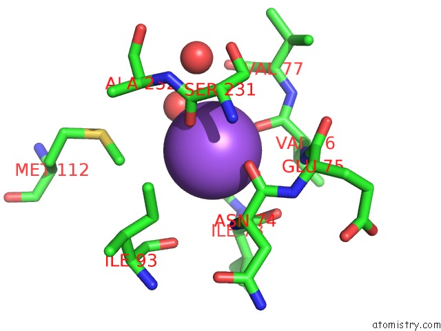
Mono view
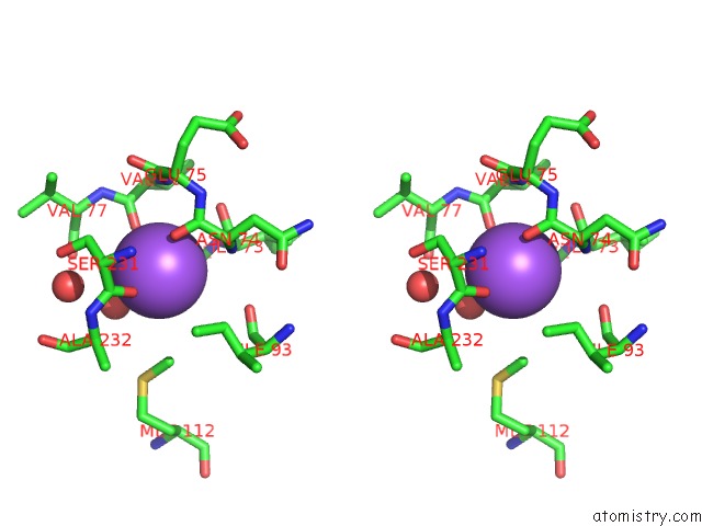
Stereo pair view
Mono view
Stereo pair view
A full contact list of Sodium with other atoms in the Na binding
site number 1 of E. Coli Methionine Aminopeptidase: Methionine Phosphonate Complex within 5.0Å range:
|
Reference:
W.T.Lowther,
Y.Zhang,
P.B.Sampson,
J.F.Honek,
B.W.Matthews.
Insights Into the Mechanism of Escherichia Coli Methionine Aminopeptidase From the Structural Analysis of Reaction Products and Phosphorus-Based Transition-State Analogues. Biochemistry V. 38 14810 1999.
ISSN: ISSN 0006-2960
PubMed: 10555963
DOI: 10.1021/BI991711G
Page generated: Sun Oct 6 17:57:45 2024
ISSN: ISSN 0006-2960
PubMed: 10555963
DOI: 10.1021/BI991711G
Last articles
Zn in 9MJ5Zn in 9HNW
Zn in 9G0L
Zn in 9FNE
Zn in 9DZN
Zn in 9E0I
Zn in 9D32
Zn in 9DAK
Zn in 8ZXC
Zn in 8ZUF