Sodium in PDB 8z2x: Crystal Structure of Exo-Beta-(1,3)-Glucanase From Aspergillus Oryzae (Aobgl) As A Complex with Cellobiose
Enzymatic activity of Crystal Structure of Exo-Beta-(1,3)-Glucanase From Aspergillus Oryzae (Aobgl) As A Complex with Cellobiose
All present enzymatic activity of Crystal Structure of Exo-Beta-(1,3)-Glucanase From Aspergillus Oryzae (Aobgl) As A Complex with Cellobiose:
3.2.1.58;
3.2.1.58;
Protein crystallography data
The structure of Crystal Structure of Exo-Beta-(1,3)-Glucanase From Aspergillus Oryzae (Aobgl) As A Complex with Cellobiose, PDB code: 8z2x
was solved by
B.Banerjee,
C.K.Kamale,
A.B.Suryawanshi,
P.Bhaumik,
with X-Ray Crystallography technique. A brief refinement statistics is given in the table below:
| Resolution Low / High (Å) | 35.03 / 1.73 |
| Space group | P 32 2 1 |
| Cell size a, b, c (Å), α, β, γ (°) | 80.89, 80.89, 113.69, 90, 90, 120 |
| R / Rfree (%) | 13.7 / 15.4 |
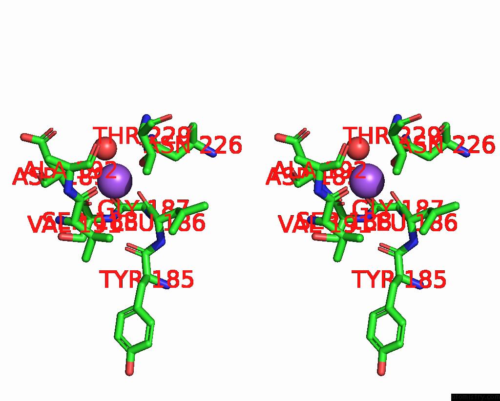
Sodium Binding Sites:
The binding sites of Sodium atom in the Crystal Structure of Exo-Beta-(1,3)-Glucanase From Aspergillus Oryzae (Aobgl) As A Complex with Cellobiose
(pdb code 8z2x). This binding sites where shown within
5.0 Angstroms radius around Sodium atom.
In total only one binding site of Sodium was determined in the Crystal Structure of Exo-Beta-(1,3)-Glucanase From Aspergillus Oryzae (Aobgl) As A Complex with Cellobiose, PDB code: 8z2x:
In total only one binding site of Sodium was determined in the Crystal Structure of Exo-Beta-(1,3)-Glucanase From Aspergillus Oryzae (Aobgl) As A Complex with Cellobiose, PDB code: 8z2x:
Sodium binding site 1 out of 1 in 8z2x
Go back to
Sodium binding site 1 out
of 1 in the Crystal Structure of Exo-Beta-(1,3)-Glucanase From Aspergillus Oryzae (Aobgl) As A Complex with Cellobiose

Mono view
Stereo pair view
Mono view
Stereo pair view
A full contact list of Sodium with other atoms in the Na binding
site number 1 of Crystal Structure of Exo-Beta-(1,3)-Glucanase From Aspergillus Oryzae (Aobgl) As A Complex with Cellobiose within 5.0Å range:
|
Reference:
B.Banerjee,
C.K.Kamale,
A.B.Suryawanshi,
S.Dasgupta,
S.Noronha,
P.Bhaumik.
Crystal Structures of Aspergillus Oryzae Exo-Beta-(1,3)-Glucanase Reveal Insights Into Oligosaccharide Binding, Recognition, and Hydrolysis. Febs Lett. 2024.
ISSN: ISSN 0014-5793
PubMed: 39448541
DOI: 10.1002/1873-3468.15045
Page generated: Wed Nov 13 13:10:36 2024
ISSN: ISSN 0014-5793
PubMed: 39448541
DOI: 10.1002/1873-3468.15045
Last articles
Zn in 9MJ5Zn in 9HNW
Zn in 9G0L
Zn in 9FNE
Zn in 9DZN
Zn in 9E0I
Zn in 9D32
Zn in 9DAK
Zn in 8ZXC
Zn in 8ZUF