Sodium in PDB 8vh7: Crystal Structure of Heparosan Synthase 2 From Pasteurella Multocida at 1.98 A
Protein crystallography data
The structure of Crystal Structure of Heparosan Synthase 2 From Pasteurella Multocida at 1.98 A, PDB code: 8vh7
was solved by
L.C.Pedersen,
J.Liu,
E.Stancanelli,
J.M.Krahn,
with X-Ray Crystallography technique. A brief refinement statistics is given in the table below:
Resolution Low / High (Å)
|
45.70 /
1.98
|
Space group
|
P 21 21 2
|
Cell size a, b, c (Å), α, β, γ (°)
|
93.178,
163.177,
84.273,
90,
90,
90
|
R / Rfree (%)
|
20.8 /
24.9
|
Other elements in 8vh7:
The structure of Crystal Structure of Heparosan Synthase 2 From Pasteurella Multocida at 1.98 A also contains other interesting chemical elements:
Sodium Binding Sites:
The binding sites of Sodium atom in the Crystal Structure of Heparosan Synthase 2 From Pasteurella Multocida at 1.98 A
(pdb code 8vh7). This binding sites where shown within
5.0 Angstroms radius around Sodium atom.
In total 4 binding sites of Sodium where determined in the
Crystal Structure of Heparosan Synthase 2 From Pasteurella Multocida at 1.98 A, PDB code: 8vh7:
Jump to Sodium binding site number:
1;
2;
3;
4;
Sodium binding site 1 out
of 4 in 8vh7
Go back to
Sodium Binding Sites List in 8vh7
Sodium binding site 1 out
of 4 in the Crystal Structure of Heparosan Synthase 2 From Pasteurella Multocida at 1.98 A
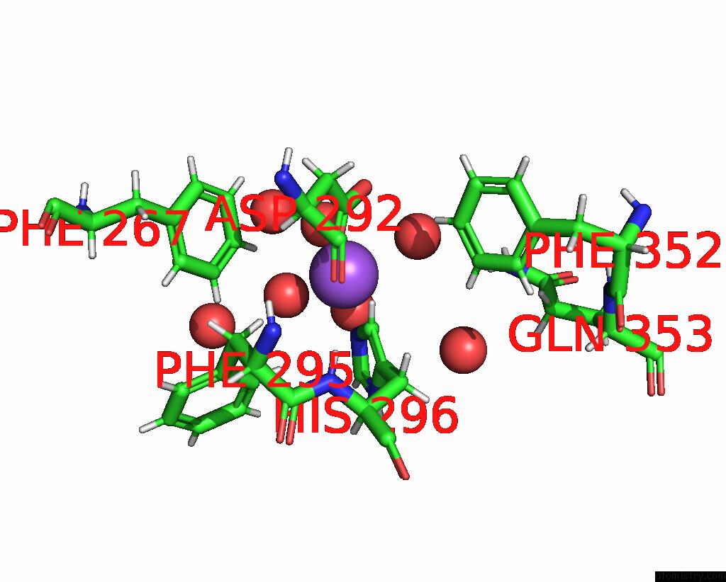
Mono view
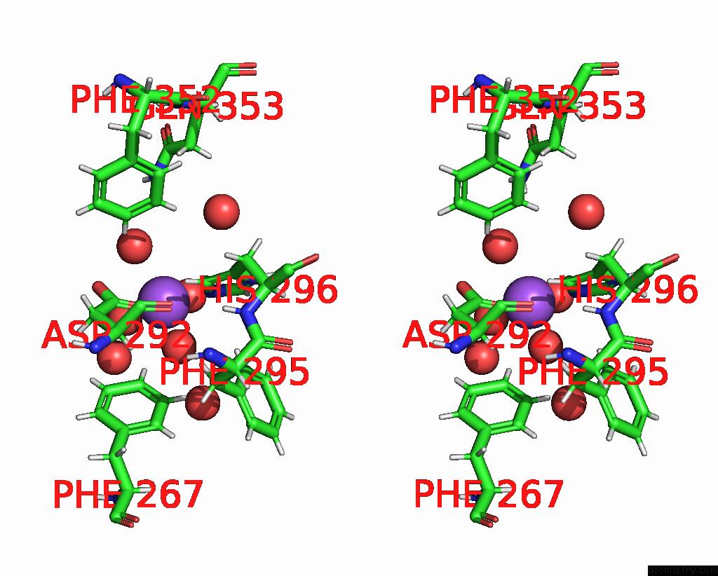
Stereo pair view
|
A full contact list of Sodium with other atoms in the Na binding
site number 1 of Crystal Structure of Heparosan Synthase 2 From Pasteurella Multocida at 1.98 A within 5.0Å range:
probe
|
atom
|
residue
|
distance (Å)
|
B
|
Occ
|
A:Na707
b:27.2
occ:1.00
|
O
|
A:HOH1123
|
2.2
|
45.4
|
1.0
|
NE2
|
A:HIS296
|
2.4
|
35.3
|
1.0
|
OD1
|
A:ASP292
|
2.4
|
26.4
|
1.0
|
O
|
A:HOH1187
|
2.5
|
46.8
|
1.0
|
O
|
A:HOH1182
|
2.7
|
49.5
|
1.0
|
O
|
A:HOH976
|
2.8
|
43.5
|
1.0
|
CG
|
A:ASP292
|
3.2
|
23.6
|
1.0
|
HZ
|
A:PHE267
|
3.2
|
25.6
|
1.0
|
CD2
|
A:HIS296
|
3.2
|
32.1
|
1.0
|
OD2
|
A:ASP292
|
3.2
|
32.7
|
1.0
|
HD2
|
A:HIS296
|
3.2
|
38.5
|
1.0
|
CE1
|
A:HIS296
|
3.4
|
31.1
|
1.0
|
HE1
|
A:HIS296
|
3.7
|
37.4
|
1.0
|
CZ
|
A:PHE267
|
4.0
|
21.3
|
1.0
|
O
|
A:HOH1237
|
4.0
|
44.6
|
1.0
|
CG
|
A:HIS296
|
4.4
|
35.4
|
1.0
|
ND1
|
A:HIS296
|
4.5
|
36.1
|
1.0
|
HE1
|
A:PHE267
|
4.6
|
26.2
|
1.0
|
CB
|
A:ASP292
|
4.6
|
21.2
|
1.0
|
HZ
|
A:PHE352
|
4.7
|
30.8
|
1.0
|
O
|
A:HOH1156
|
4.7
|
47.4
|
1.0
|
CE1
|
A:PHE267
|
4.7
|
21.8
|
1.0
|
HE2
|
A:PHE267
|
4.7
|
27.1
|
1.0
|
HA
|
A:ASP292
|
4.7
|
21.9
|
1.0
|
HD2
|
A:PHE295
|
4.8
|
27.3
|
1.0
|
CE2
|
A:PHE267
|
4.8
|
22.6
|
1.0
|
HE21
|
A:GLN353
|
4.9
|
57.5
|
1.0
|
O
|
A:HOH1107
|
4.9
|
37.6
|
1.0
|
HB2
|
A:ASP292
|
4.9
|
25.5
|
1.0
|
O
|
A:ASP292
|
5.0
|
17.9
|
1.0
|
|
Sodium binding site 2 out
of 4 in 8vh7
Go back to
Sodium Binding Sites List in 8vh7
Sodium binding site 2 out
of 4 in the Crystal Structure of Heparosan Synthase 2 From Pasteurella Multocida at 1.98 A
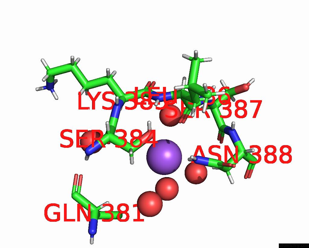
Mono view
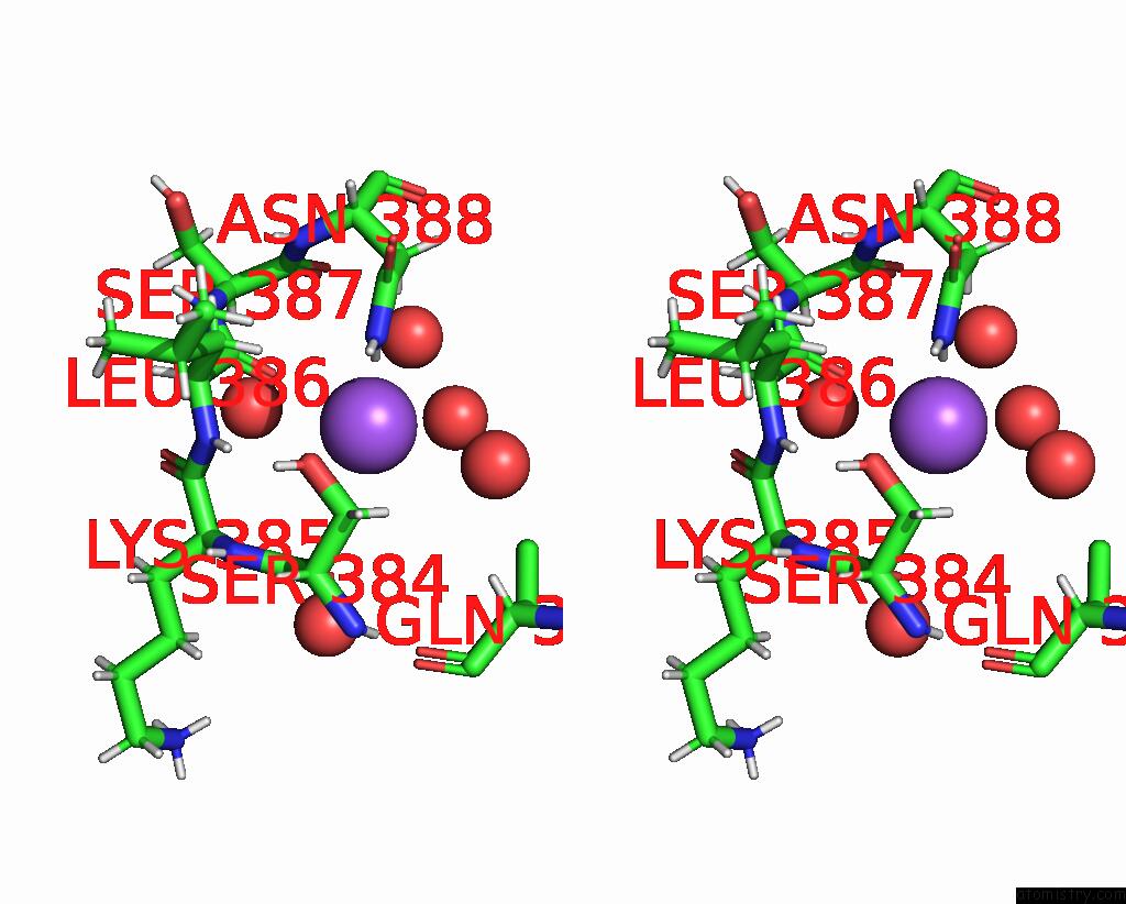
Stereo pair view
|
A full contact list of Sodium with other atoms in the Na binding
site number 2 of Crystal Structure of Heparosan Synthase 2 From Pasteurella Multocida at 1.98 A within 5.0Å range:
probe
|
atom
|
residue
|
distance (Å)
|
B
|
Occ
|
A:Na708
b:44.0
occ:1.00
|
HD22
|
A:ASN388
|
2.2
|
29.8
|
1.0
|
O
|
A:HOH1209
|
2.3
|
44.6
|
1.0
|
O
|
A:LEU386
|
2.6
|
17.9
|
1.0
|
ND2
|
A:ASN388
|
3.0
|
24.9
|
1.0
|
HB2
|
A:ASN388
|
3.3
|
28.9
|
1.0
|
HD21
|
A:ASN388
|
3.4
|
29.8
|
1.0
|
O
|
A:HOH957
|
3.5
|
50.0
|
1.0
|
C
|
A:LEU386
|
3.7
|
21.5
|
1.0
|
O
|
A:SER384
|
3.9
|
20.6
|
1.0
|
CG
|
A:ASN388
|
4.0
|
25.8
|
1.0
|
CB
|
A:ASN388
|
4.1
|
24.1
|
1.0
|
HB3
|
A:SER384
|
4.1
|
23.8
|
1.0
|
N
|
A:LEU386
|
4.1
|
17.9
|
1.0
|
OG
|
A:SER384
|
4.2
|
22.5
|
1.0
|
H
|
A:LEU386
|
4.2
|
21.4
|
1.0
|
HA
|
A:LYS385
|
4.2
|
22.6
|
1.0
|
O
|
A:HOH964
|
4.3
|
34.3
|
1.0
|
HB2
|
A:LEU386
|
4.3
|
22.1
|
1.0
|
C
|
A:LYS385
|
4.4
|
17.6
|
1.0
|
C
|
A:SER384
|
4.4
|
18.8
|
1.0
|
O
|
A:SER387
|
4.5
|
18.9
|
1.0
|
CA
|
A:LEU386
|
4.5
|
21.1
|
1.0
|
C
|
A:SER387
|
4.5
|
21.8
|
1.0
|
HA
|
A:SER387
|
4.6
|
28.5
|
1.0
|
CB
|
A:SER384
|
4.6
|
19.8
|
1.0
|
HB3
|
A:ASN388
|
4.7
|
28.9
|
1.0
|
CA
|
A:LYS385
|
4.7
|
18.8
|
1.0
|
N
|
A:ASN388
|
4.7
|
18.7
|
1.0
|
N
|
A:SER387
|
4.7
|
19.3
|
1.0
|
O
|
A:HOH1077
|
4.8
|
27.0
|
1.0
|
HA
|
A:GLN381
|
4.8
|
37.0
|
1.0
|
N
|
A:LYS385
|
4.8
|
19.9
|
1.0
|
HG
|
A:SER384
|
4.8
|
27.0
|
1.0
|
CA
|
A:SER387
|
4.9
|
23.7
|
1.0
|
O
|
A:LYS385
|
4.9
|
19.0
|
1.0
|
CG
|
A:GLN381
|
4.9
|
53.8
|
1.0
|
CB
|
A:LEU386
|
4.9
|
18.4
|
1.0
|
H
|
A:ASN388
|
5.0
|
22.5
|
1.0
|
O
|
A:HOH933
|
5.0
|
38.0
|
1.0
|
|
Sodium binding site 3 out
of 4 in 8vh7
Go back to
Sodium Binding Sites List in 8vh7
Sodium binding site 3 out
of 4 in the Crystal Structure of Heparosan Synthase 2 From Pasteurella Multocida at 1.98 A
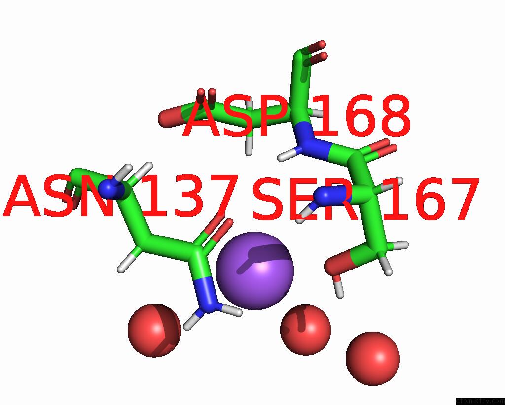
Mono view
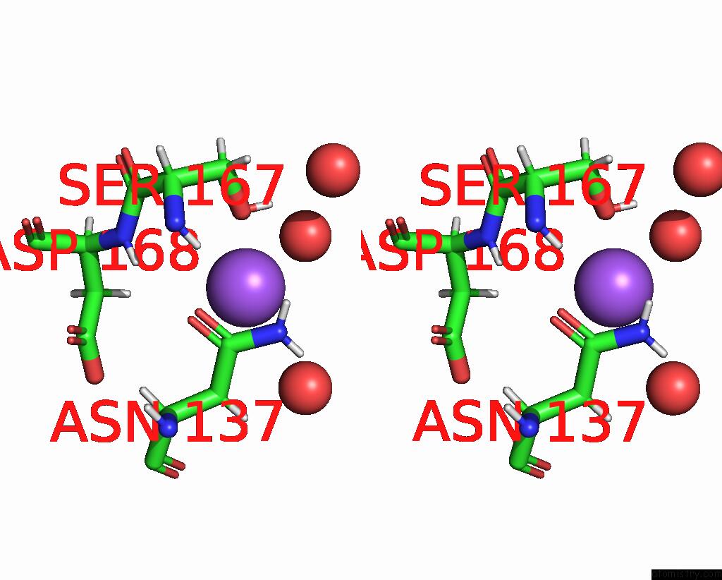
Stereo pair view
|
A full contact list of Sodium with other atoms in the Na binding
site number 3 of Crystal Structure of Heparosan Synthase 2 From Pasteurella Multocida at 1.98 A within 5.0Å range:
probe
|
atom
|
residue
|
distance (Å)
|
B
|
Occ
|
A:Na709
b:58.5
occ:1.00
|
O
|
A:HOH1232
|
2.7
|
61.6
|
1.0
|
O
|
A:HOH1191
|
3.4
|
47.8
|
1.0
|
HB3
|
A:ASP168
|
3.5
|
31.9
|
1.0
|
HG
|
A:SER167
|
3.5
|
47.6
|
1.0
|
OG
|
A:SER167
|
3.6
|
39.6
|
1.0
|
HB3
|
A:ASN137
|
3.7
|
26.0
|
1.0
|
CG
|
A:ASN137
|
4.0
|
24.6
|
1.0
|
ND2
|
A:ASN137
|
4.0
|
24.1
|
1.0
|
HD22
|
A:ASN137
|
4.2
|
28.9
|
1.0
|
HD21
|
A:ASN137
|
4.2
|
28.9
|
1.0
|
O
|
A:HOH1184
|
4.2
|
50.7
|
1.0
|
OD1
|
A:ASN137
|
4.3
|
23.9
|
1.0
|
CB
|
A:ASN137
|
4.3
|
21.7
|
1.0
|
H
|
A:ASP168
|
4.4
|
24.6
|
1.0
|
CB
|
A:ASP168
|
4.4
|
26.6
|
1.0
|
N
|
A:ASP168
|
4.6
|
20.5
|
1.0
|
HB2
|
A:ASN137
|
4.9
|
26.0
|
1.0
|
CB
|
A:SER167
|
4.9
|
29.1
|
1.0
|
HB2
|
A:ASP168
|
5.0
|
31.9
|
1.0
|
CA
|
A:ASP168
|
5.0
|
28.0
|
1.0
|
HA
|
A:ASP168
|
5.0
|
33.6
|
1.0
|
|
Sodium binding site 4 out
of 4 in 8vh7
Go back to
Sodium Binding Sites List in 8vh7
Sodium binding site 4 out
of 4 in the Crystal Structure of Heparosan Synthase 2 From Pasteurella Multocida at 1.98 A
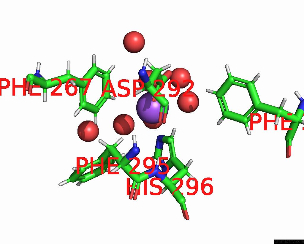
Mono view
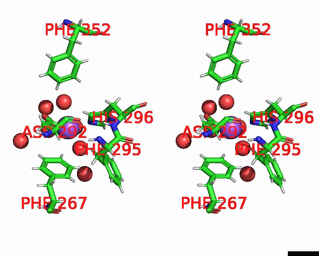
Stereo pair view
|
A full contact list of Sodium with other atoms in the Na binding
site number 4 of Crystal Structure of Heparosan Synthase 2 From Pasteurella Multocida at 1.98 A within 5.0Å range:
probe
|
atom
|
residue
|
distance (Å)
|
B
|
Occ
|
B:Na705
b:25.2
occ:1.00
|
O
|
B:HOH1076
|
2.2
|
41.8
|
1.0
|
OD1
|
B:ASP292
|
2.4
|
30.4
|
1.0
|
NE2
|
B:HIS296
|
2.4
|
32.0
|
1.0
|
O
|
B:HOH1164
|
2.5
|
41.0
|
1.0
|
O
|
B:HOH1140
|
2.5
|
34.0
|
1.0
|
O
|
B:HOH1068
|
2.6
|
32.9
|
1.0
|
OD2
|
B:ASP292
|
3.1
|
33.4
|
1.0
|
CG
|
B:ASP292
|
3.1
|
23.0
|
1.0
|
HZ
|
B:PHE267
|
3.1
|
30.0
|
1.0
|
CD2
|
B:HIS296
|
3.3
|
23.7
|
1.0
|
HD2
|
B:HIS296
|
3.4
|
28.4
|
1.0
|
CE1
|
B:HIS296
|
3.5
|
31.7
|
1.0
|
HE1
|
B:HIS296
|
3.6
|
38.0
|
1.0
|
O
|
B:HOH1169
|
3.7
|
50.2
|
1.0
|
CZ
|
B:PHE267
|
3.9
|
25.0
|
1.0
|
HE2
|
B:PHE267
|
4.5
|
29.3
|
1.0
|
CB
|
B:ASP292
|
4.5
|
22.6
|
1.0
|
CG
|
B:HIS296
|
4.5
|
29.5
|
1.0
|
ND1
|
B:HIS296
|
4.6
|
34.3
|
1.0
|
CE2
|
B:PHE267
|
4.6
|
24.4
|
1.0
|
HD2
|
B:PHE295
|
4.7
|
27.4
|
1.0
|
HE1
|
B:PHE267
|
4.7
|
30.6
|
1.0
|
HA
|
B:ASP292
|
4.8
|
28.6
|
1.0
|
CE1
|
B:PHE267
|
4.8
|
25.5
|
1.0
|
HB2
|
B:ASP292
|
4.8
|
27.1
|
1.0
|
HZ
|
B:PHE352
|
4.9
|
36.5
|
1.0
|
O
|
B:HOH1168
|
4.9
|
50.2
|
1.0
|
O
|
B:HOH1119
|
5.0
|
41.5
|
1.0
|
|
Reference:
E.Stancanelli,
J.A.Krahn,
E.Viverette,
R.Dutcher,
V.Pagadala,
M.J.Borgnia,
J.Liu,
L.C.Pedersen.
Structural and Functional Analysis of Heparosan Synthase 2 From Pasteurella Multocida to Improve the Synthesis of Heparin Acs Catalysis V. 14 6577 2024.
ISSN: ESSN 2155-5435
DOI: 10.1021/ACSCATAL.4C00677
Page generated: Wed Oct 9 14:01:17 2024
|