Sodium in PDB 8rwl: Crystal Structure of Methanopyrus Kandleri Malate Dehydrogenase Mutant 1
Enzymatic activity of Crystal Structure of Methanopyrus Kandleri Malate Dehydrogenase Mutant 1
All present enzymatic activity of Crystal Structure of Methanopyrus Kandleri Malate Dehydrogenase Mutant 1:
1.1.1.299;
1.1.1.299;
Protein crystallography data
The structure of Crystal Structure of Methanopyrus Kandleri Malate Dehydrogenase Mutant 1, PDB code: 8rwl
was solved by
S.Coquille,
J.Roche,
S.Engilberge,
E.Girard,
D.Madern,
with X-Ray Crystallography technique. A brief refinement statistics is given in the table below:
| Resolution Low / High (Å) | 19.92 / 2.30 |
| Space group | P 43 21 2 |
| Cell size a, b, c (Å), α, β, γ (°) | 78.25, 78.25, 251.3, 90, 90, 90 |
| R / Rfree (%) | 20.4 / 24.3 |
Other elements in 8rwl:
The structure of Crystal Structure of Methanopyrus Kandleri Malate Dehydrogenase Mutant 1 also contains other interesting chemical elements:
| Chlorine | (Cl) | 3 atoms |
Sodium Binding Sites:
The binding sites of Sodium atom in the Crystal Structure of Methanopyrus Kandleri Malate Dehydrogenase Mutant 1
(pdb code 8rwl). This binding sites where shown within
5.0 Angstroms radius around Sodium atom.
In total 2 binding sites of Sodium where determined in the Crystal Structure of Methanopyrus Kandleri Malate Dehydrogenase Mutant 1, PDB code: 8rwl:
Jump to Sodium binding site number: 1; 2;
In total 2 binding sites of Sodium where determined in the Crystal Structure of Methanopyrus Kandleri Malate Dehydrogenase Mutant 1, PDB code: 8rwl:
Jump to Sodium binding site number: 1; 2;
Sodium binding site 1 out of 2 in 8rwl
Go back to
Sodium binding site 1 out
of 2 in the Crystal Structure of Methanopyrus Kandleri Malate Dehydrogenase Mutant 1
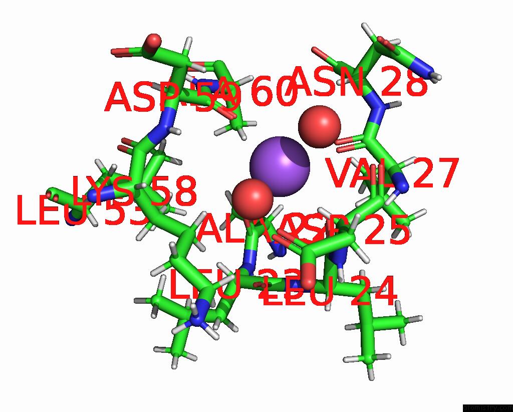
Mono view
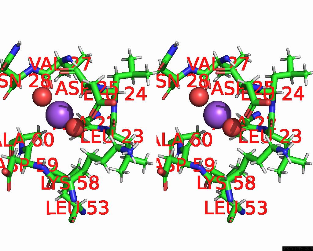
Stereo pair view
Mono view
Stereo pair view
A full contact list of Sodium with other atoms in the Na binding
site number 1 of Crystal Structure of Methanopyrus Kandleri Malate Dehydrogenase Mutant 1 within 5.0Å range:
|
Sodium binding site 2 out of 2 in 8rwl
Go back to
Sodium binding site 2 out
of 2 in the Crystal Structure of Methanopyrus Kandleri Malate Dehydrogenase Mutant 1
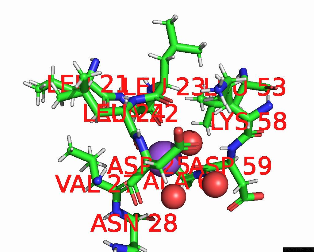
Mono view
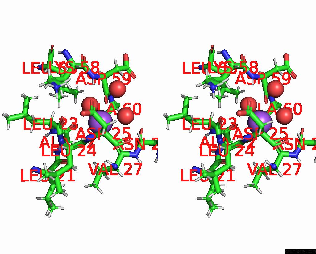
Stereo pair view
Mono view
Stereo pair view
A full contact list of Sodium with other atoms in the Na binding
site number 2 of Crystal Structure of Methanopyrus Kandleri Malate Dehydrogenase Mutant 1 within 5.0Å range:
|
Reference:
S.Coquille,
C.Simoes Pereira,
C.Brochier-Armanet,
J.Roche,
G.Santoni,
N.Coquelle,
E.Girard,
F.Sterpone,
D.Madern.
Navigating the Conformational Landscape of An Enzyme. Stabilization of A Low Populated Conformer By Evolutionary Mutations Triggers Allostery Into A Non-Allosteric Enzyme. To Be Published.
Page generated: Wed Oct 9 13:23:53 2024
Last articles
Zn in 9J0NZn in 9J0O
Zn in 9J0P
Zn in 9FJX
Zn in 9EKB
Zn in 9C0F
Zn in 9CAH
Zn in 9CH0
Zn in 9CH3
Zn in 9CH1