Sodium in PDB 8owm: Crystal Structure of Glutamate Dehydrogenase 2 From Arabidopsis Thaliana Binding Ca, Nad and 2,2-Dihydroxyglutarate
Enzymatic activity of Crystal Structure of Glutamate Dehydrogenase 2 From Arabidopsis Thaliana Binding Ca, Nad and 2,2-Dihydroxyglutarate
All present enzymatic activity of Crystal Structure of Glutamate Dehydrogenase 2 From Arabidopsis Thaliana Binding Ca, Nad and 2,2-Dihydroxyglutarate:
1.4.1.3;
1.4.1.3;
Protein crystallography data
The structure of Crystal Structure of Glutamate Dehydrogenase 2 From Arabidopsis Thaliana Binding Ca, Nad and 2,2-Dihydroxyglutarate, PDB code: 8owm
was solved by
M.Grzechowiak,
M.Ruszkowski,
with X-Ray Crystallography technique. A brief refinement statistics is given in the table below:
| Resolution Low / High (Å) | 65.70 / 1.70 |
| Space group | P 1 |
| Cell size a, b, c (Å), α, β, γ (°) | 95.544, 95.629, 95.841, 90.42, 93.59, 117.78 |
| R / Rfree (%) | 14.4 / 17.2 |
Other elements in 8owm:
The structure of Crystal Structure of Glutamate Dehydrogenase 2 From Arabidopsis Thaliana Binding Ca, Nad and 2,2-Dihydroxyglutarate also contains other interesting chemical elements:
| Calcium | (Ca) | 6 atoms |
Sodium Binding Sites:
The binding sites of Sodium atom in the Crystal Structure of Glutamate Dehydrogenase 2 From Arabidopsis Thaliana Binding Ca, Nad and 2,2-Dihydroxyglutarate
(pdb code 8owm). This binding sites where shown within
5.0 Angstroms radius around Sodium atom.
In total 3 binding sites of Sodium where determined in the Crystal Structure of Glutamate Dehydrogenase 2 From Arabidopsis Thaliana Binding Ca, Nad and 2,2-Dihydroxyglutarate, PDB code: 8owm:
Jump to Sodium binding site number: 1; 2; 3;
In total 3 binding sites of Sodium where determined in the Crystal Structure of Glutamate Dehydrogenase 2 From Arabidopsis Thaliana Binding Ca, Nad and 2,2-Dihydroxyglutarate, PDB code: 8owm:
Jump to Sodium binding site number: 1; 2; 3;
Sodium binding site 1 out of 3 in 8owm
Go back to
Sodium binding site 1 out
of 3 in the Crystal Structure of Glutamate Dehydrogenase 2 From Arabidopsis Thaliana Binding Ca, Nad and 2,2-Dihydroxyglutarate

Mono view
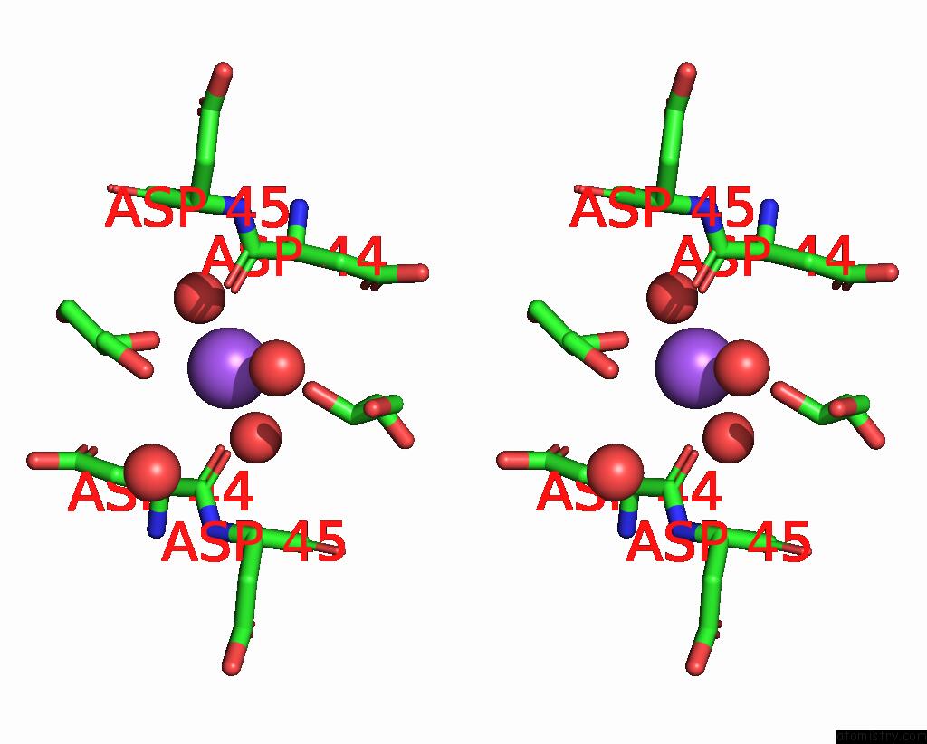
Stereo pair view
Mono view
Stereo pair view
A full contact list of Sodium with other atoms in the Na binding
site number 1 of Crystal Structure of Glutamate Dehydrogenase 2 From Arabidopsis Thaliana Binding Ca, Nad and 2,2-Dihydroxyglutarate within 5.0Å range:
|
Sodium binding site 2 out of 3 in 8owm
Go back to
Sodium binding site 2 out
of 3 in the Crystal Structure of Glutamate Dehydrogenase 2 From Arabidopsis Thaliana Binding Ca, Nad and 2,2-Dihydroxyglutarate
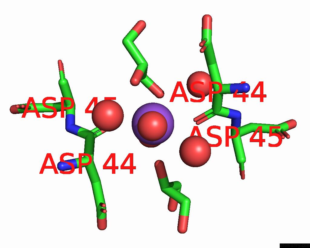
Mono view

Stereo pair view
Mono view
Stereo pair view
A full contact list of Sodium with other atoms in the Na binding
site number 2 of Crystal Structure of Glutamate Dehydrogenase 2 From Arabidopsis Thaliana Binding Ca, Nad and 2,2-Dihydroxyglutarate within 5.0Å range:
|
Sodium binding site 3 out of 3 in 8owm
Go back to
Sodium binding site 3 out
of 3 in the Crystal Structure of Glutamate Dehydrogenase 2 From Arabidopsis Thaliana Binding Ca, Nad and 2,2-Dihydroxyglutarate
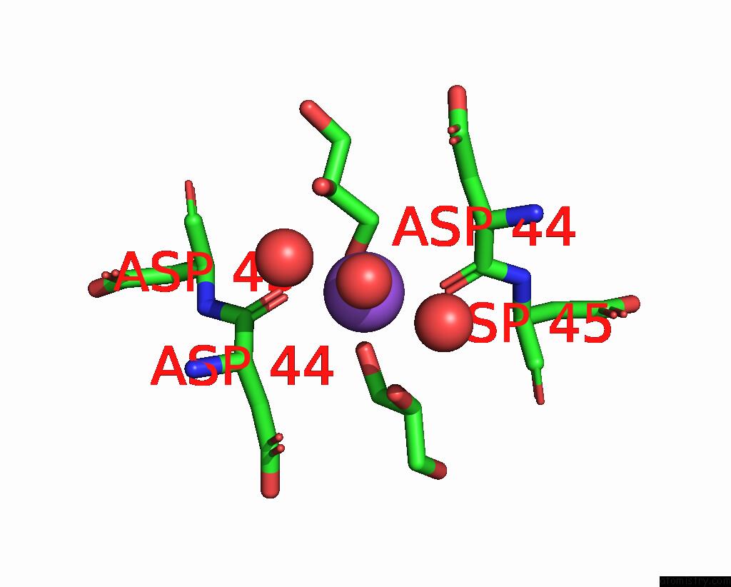
Mono view
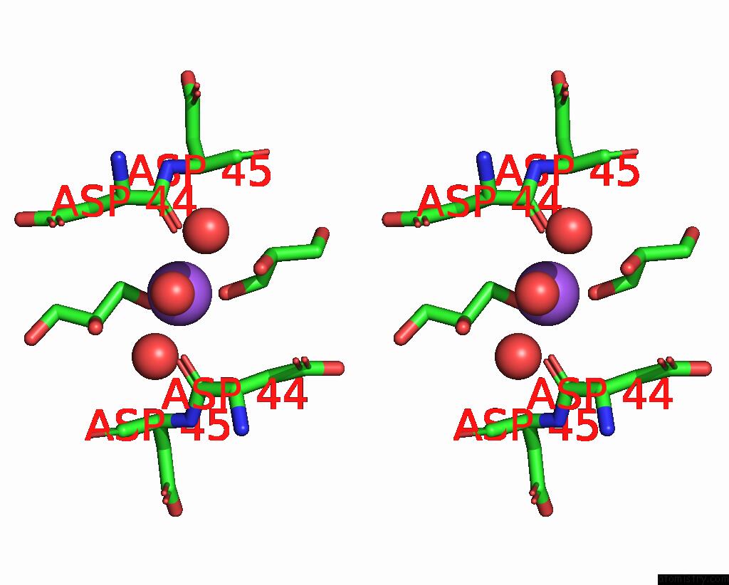
Stereo pair view
Mono view
Stereo pair view
A full contact list of Sodium with other atoms in the Na binding
site number 3 of Crystal Structure of Glutamate Dehydrogenase 2 From Arabidopsis Thaliana Binding Ca, Nad and 2,2-Dihydroxyglutarate within 5.0Å range:
|
Reference:
M.Grzechowiak,
J.Sliwiak,
M.Jaskolski,
M.Ruszkowski.
Structural and Functional Studies of Arabidopsis Thaliana Glutamate Dehydrogenase Isoform 2 Demonstrate Enzyme Dynamics and Identify Its Calcium Binding Site. Plant Physiol Biochem. V. 201 07895 2023.
ISSN: ESSN 1873-2690
PubMed: 37478728
DOI: 10.1016/J.PLAPHY.2023.107895
Page generated: Wed Oct 9 12:49:09 2024
ISSN: ESSN 1873-2690
PubMed: 37478728
DOI: 10.1016/J.PLAPHY.2023.107895
Last articles
Zn in 9J0NZn in 9J0O
Zn in 9J0P
Zn in 9FJX
Zn in 9EKB
Zn in 9C0F
Zn in 9CAH
Zn in 9CH0
Zn in 9CH3
Zn in 9CH1